Magnesium »
PDB 6cgf-6cq2 »
6cj4 »
Magnesium in PDB 6cj4: Crystal Structure of Protein Cite From Mycobacterium Tuberculosis in Complex with Magnesium and Acetoacetate
Protein crystallography data
The structure of Crystal Structure of Protein Cite From Mycobacterium Tuberculosis in Complex with Magnesium and Acetoacetate, PDB code: 6cj4
was solved by
A.A.Fedorov,
E.V.Fedorov,
H.Wang,
J.B.Bonanno,
L.Carvalho,
S.C.Almo,
with X-Ray Crystallography technique. A brief refinement statistics is given in the table below:
| Resolution Low / High (Å) | 25.00 / 1.73 |
| Space group | H 3 2 |
| Cell size a, b, c (Å), α, β, γ (°) | 91.673, 91.673, 221.386, 90.00, 90.00, 120.00 |
| R / Rfree (%) | 18.3 / 20.2 |
Magnesium Binding Sites:
The binding sites of Magnesium atom in the Crystal Structure of Protein Cite From Mycobacterium Tuberculosis in Complex with Magnesium and Acetoacetate
(pdb code 6cj4). This binding sites where shown within
5.0 Angstroms radius around Magnesium atom.
In total only one binding site of Magnesium was determined in the Crystal Structure of Protein Cite From Mycobacterium Tuberculosis in Complex with Magnesium and Acetoacetate, PDB code: 6cj4:
In total only one binding site of Magnesium was determined in the Crystal Structure of Protein Cite From Mycobacterium Tuberculosis in Complex with Magnesium and Acetoacetate, PDB code: 6cj4:
Magnesium binding site 1 out of 1 in 6cj4
Go back to
Magnesium binding site 1 out
of 1 in the Crystal Structure of Protein Cite From Mycobacterium Tuberculosis in Complex with Magnesium and Acetoacetate
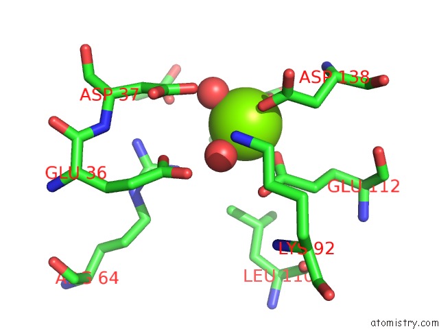
Mono view
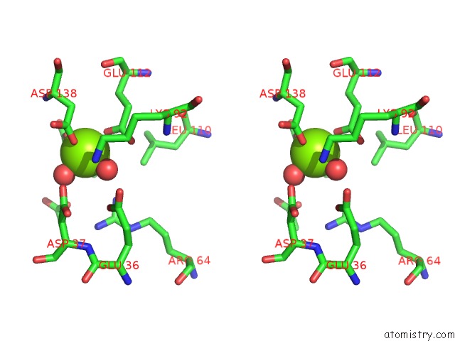
Stereo pair view
Mono view
Stereo pair view
A full contact list of Magnesium with other atoms in the Mg binding
site number 1 of Crystal Structure of Protein Cite From Mycobacterium Tuberculosis in Complex with Magnesium and Acetoacetate within 5.0Å range:
|
Reference:
H.Wang,
A.A.Fedorov,
E.V.Fedorov,
D.M.Hunt,
A.Rodgers,
H.L.Douglas,
A.Garza-Garcia,
J.B.Bonanno,
S.C.Almo,
L.P.S.De Carvalho.
An Essential Bifunctional Enzyme Inmycobacterium Tuberculosisfor Itaconate Dissimilation and Leucine Catabolism. Proc.Natl.Acad.Sci.Usa V. 116 15907 2019.
ISSN: ESSN 1091-6490
PubMed: 31320588
DOI: 10.1073/PNAS.1906606116
Page generated: Wed Aug 13 04:36:18 2025
ISSN: ESSN 1091-6490
PubMed: 31320588
DOI: 10.1073/PNAS.1906606116
Last articles
Mg in 7BLXMg in 7BLZ
Mg in 7BOD
Mg in 7BNR
Mg in 7BNK
Mg in 7BMC
Mg in 7BM9
Mg in 7BM8
Mg in 7BM6
Mg in 7BL4