Magnesium »
PDB 6cgf-6cq2 »
6cn1 »
Magnesium in PDB 6cn1: 2.75 Angstrom Resolution Crystal Structure of Udp-N-Acetylglucosamine 1-Carboxyvinyltransferase From Pseudomonas Putida in Complex with Uridine-Diphosphate-2(N-Acetylglucosaminyl) Butyric Acid, (2R)-2- (Phosphonooxy)Propanoic Acid and Magnesium
Enzymatic activity of 2.75 Angstrom Resolution Crystal Structure of Udp-N-Acetylglucosamine 1-Carboxyvinyltransferase From Pseudomonas Putida in Complex with Uridine-Diphosphate-2(N-Acetylglucosaminyl) Butyric Acid, (2R)-2- (Phosphonooxy)Propanoic Acid and Magnesium
All present enzymatic activity of 2.75 Angstrom Resolution Crystal Structure of Udp-N-Acetylglucosamine 1-Carboxyvinyltransferase From Pseudomonas Putida in Complex with Uridine-Diphosphate-2(N-Acetylglucosaminyl) Butyric Acid, (2R)-2- (Phosphonooxy)Propanoic Acid and Magnesium:
2.5.1.7;
2.5.1.7;
Protein crystallography data
The structure of 2.75 Angstrom Resolution Crystal Structure of Udp-N-Acetylglucosamine 1-Carboxyvinyltransferase From Pseudomonas Putida in Complex with Uridine-Diphosphate-2(N-Acetylglucosaminyl) Butyric Acid, (2R)-2- (Phosphonooxy)Propanoic Acid and Magnesium, PDB code: 6cn1
was solved by
G.Minasov,
L.Shuvalova,
I.Dubrovska,
A.Cardona-Correa,
S.Grimshaw,
K.Kwon,
W.F.Anderson,
K.J.F.Satchell,
A.Joachimiak,
Center For Structuralgenomics Of Infectious Diseases (Csgid),
with X-Ray Crystallography technique. A brief refinement statistics is given in the table below:
| Resolution Low / High (Å) | 29.95 / 2.75 |
| Space group | P 21 21 21 |
| Cell size a, b, c (Å), α, β, γ (°) | 128.615, 148.119, 164.627, 90.00, 90.00, 90.00 |
| R / Rfree (%) | 21 / 25.2 |
Other elements in 6cn1:
The structure of 2.75 Angstrom Resolution Crystal Structure of Udp-N-Acetylglucosamine 1-Carboxyvinyltransferase From Pseudomonas Putida in Complex with Uridine-Diphosphate-2(N-Acetylglucosaminyl) Butyric Acid, (2R)-2- (Phosphonooxy)Propanoic Acid and Magnesium also contains other interesting chemical elements:
| Chlorine | (Cl) | 5 atoms |
Magnesium Binding Sites:
The binding sites of Magnesium atom in the 2.75 Angstrom Resolution Crystal Structure of Udp-N-Acetylglucosamine 1-Carboxyvinyltransferase From Pseudomonas Putida in Complex with Uridine-Diphosphate-2(N-Acetylglucosaminyl) Butyric Acid, (2R)-2- (Phosphonooxy)Propanoic Acid and Magnesium
(pdb code 6cn1). This binding sites where shown within
5.0 Angstroms radius around Magnesium atom.
In total 8 binding sites of Magnesium where determined in the 2.75 Angstrom Resolution Crystal Structure of Udp-N-Acetylglucosamine 1-Carboxyvinyltransferase From Pseudomonas Putida in Complex with Uridine-Diphosphate-2(N-Acetylglucosaminyl) Butyric Acid, (2R)-2- (Phosphonooxy)Propanoic Acid and Magnesium, PDB code: 6cn1:
Jump to Magnesium binding site number: 1; 2; 3; 4; 5; 6; 7; 8;
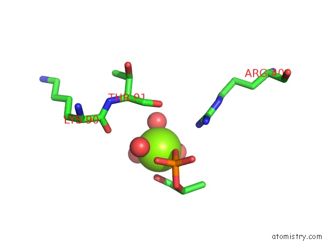
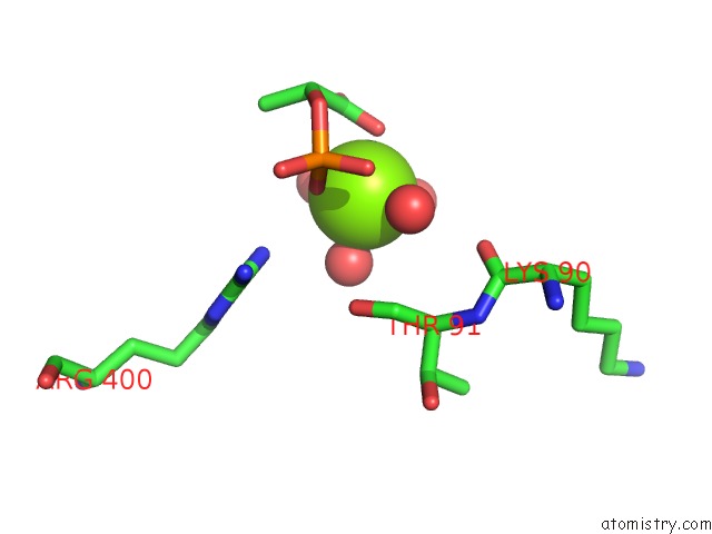
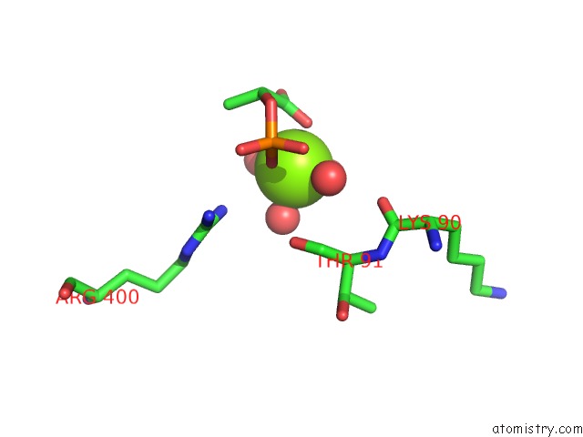
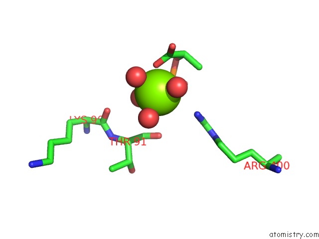
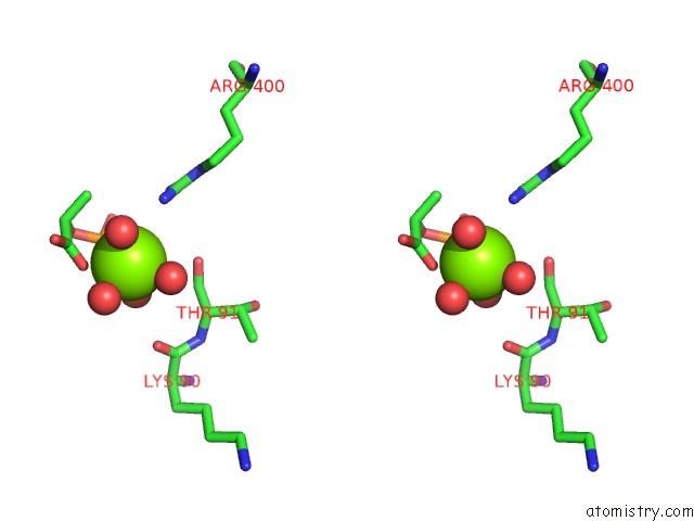
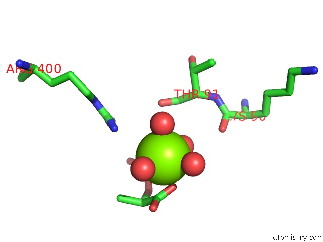
Magnesium binding site 1 out of 8 in 6cn1
Go back to
Magnesium binding site 1 out
of 8 in the 2.75 Angstrom Resolution Crystal Structure of Udp-N-Acetylglucosamine 1-Carboxyvinyltransferase From Pseudomonas Putida in Complex with Uridine-Diphosphate-2(N-Acetylglucosaminyl) Butyric Acid, (2R)-2- (Phosphonooxy)Propanoic Acid and Magnesium

Mono view

Stereo pair view
Mono view
Stereo pair view
A full contact list of Magnesium with other atoms in the Mg binding
site number 1 of 2.75 Angstrom Resolution Crystal Structure of Udp-N-Acetylglucosamine 1-Carboxyvinyltransferase From Pseudomonas Putida in Complex with Uridine-Diphosphate-2(N-Acetylglucosaminyl) Butyric Acid, (2R)-2- (Phosphonooxy)Propanoic Acid and Magnesium within 5.0Å range:
|
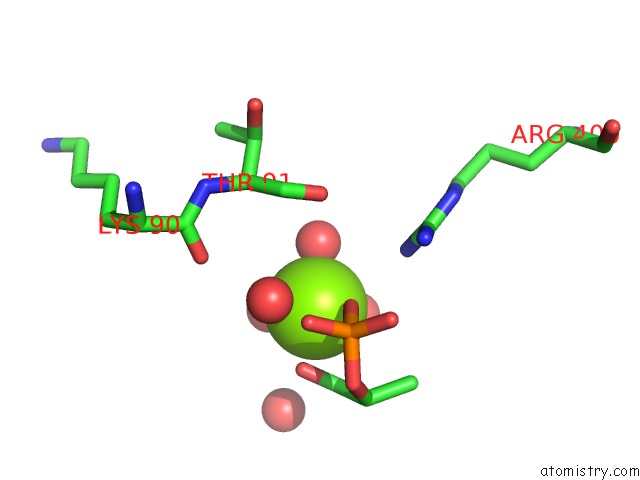
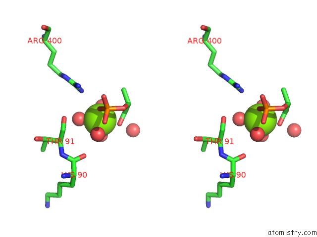
Magnesium binding site 2 out of 8 in 6cn1
Go back to
Magnesium binding site 2 out
of 8 in the 2.75 Angstrom Resolution Crystal Structure of Udp-N-Acetylglucosamine 1-Carboxyvinyltransferase From Pseudomonas Putida in Complex with Uridine-Diphosphate-2(N-Acetylglucosaminyl) Butyric Acid, (2R)-2- (Phosphonooxy)Propanoic Acid and Magnesium
Mono view

Stereo pair view
Mono view
Stereo pair view
A full contact list of Magnesium with other atoms in the Mg binding
site number 2 of 2.75 Angstrom Resolution Crystal Structure of Udp-N-Acetylglucosamine 1-Carboxyvinyltransferase From Pseudomonas Putida in Complex with Uridine-Diphosphate-2(N-Acetylglucosaminyl) Butyric Acid, (2R)-2- (Phosphonooxy)Propanoic Acid and Magnesium within 5.0Å range:
|
Magnesium binding site 3 out of 8 in 6cn1
Go back to
Magnesium binding site 3 out
of 8 in the 2.75 Angstrom Resolution Crystal Structure of Udp-N-Acetylglucosamine 1-Carboxyvinyltransferase From Pseudomonas Putida in Complex with Uridine-Diphosphate-2(N-Acetylglucosaminyl) Butyric Acid, (2R)-2- (Phosphonooxy)Propanoic Acid and Magnesium
Mono view

Stereo pair view
Mono view
Stereo pair view
A full contact list of Magnesium with other atoms in the Mg binding
site number 3 of 2.75 Angstrom Resolution Crystal Structure of Udp-N-Acetylglucosamine 1-Carboxyvinyltransferase From Pseudomonas Putida in Complex with Uridine-Diphosphate-2(N-Acetylglucosaminyl) Butyric Acid, (2R)-2- (Phosphonooxy)Propanoic Acid and Magnesium within 5.0Å range:
|
Magnesium binding site 4 out of 8 in 6cn1
Go back to
Magnesium binding site 4 out
of 8 in the 2.75 Angstrom Resolution Crystal Structure of Udp-N-Acetylglucosamine 1-Carboxyvinyltransferase From Pseudomonas Putida in Complex with Uridine-Diphosphate-2(N-Acetylglucosaminyl) Butyric Acid, (2R)-2- (Phosphonooxy)Propanoic Acid and Magnesium

Mono view

Stereo pair view
Mono view
Stereo pair view
A full contact list of Magnesium with other atoms in the Mg binding
site number 4 of 2.75 Angstrom Resolution Crystal Structure of Udp-N-Acetylglucosamine 1-Carboxyvinyltransferase From Pseudomonas Putida in Complex with Uridine-Diphosphate-2(N-Acetylglucosaminyl) Butyric Acid, (2R)-2- (Phosphonooxy)Propanoic Acid and Magnesium within 5.0Å range:
|
Magnesium binding site 5 out of 8 in 6cn1
Go back to
Magnesium binding site 5 out
of 8 in the 2.75 Angstrom Resolution Crystal Structure of Udp-N-Acetylglucosamine 1-Carboxyvinyltransferase From Pseudomonas Putida in Complex with Uridine-Diphosphate-2(N-Acetylglucosaminyl) Butyric Acid, (2R)-2- (Phosphonooxy)Propanoic Acid and Magnesium
Mono view

Stereo pair view
Mono view
Stereo pair view
A full contact list of Magnesium with other atoms in the Mg binding
site number 5 of 2.75 Angstrom Resolution Crystal Structure of Udp-N-Acetylglucosamine 1-Carboxyvinyltransferase From Pseudomonas Putida in Complex with Uridine-Diphosphate-2(N-Acetylglucosaminyl) Butyric Acid, (2R)-2- (Phosphonooxy)Propanoic Acid and Magnesium within 5.0Å range:
|
Magnesium binding site 6 out of 8 in 6cn1
Go back to
Magnesium binding site 6 out
of 8 in the 2.75 Angstrom Resolution Crystal Structure of Udp-N-Acetylglucosamine 1-Carboxyvinyltransferase From Pseudomonas Putida in Complex with Uridine-Diphosphate-2(N-Acetylglucosaminyl) Butyric Acid, (2R)-2- (Phosphonooxy)Propanoic Acid and Magnesium
Mono view
Stereo pair view
Mono view
Stereo pair view
A full contact list of Magnesium with other atoms in the Mg binding
site number 6 of 2.75 Angstrom Resolution Crystal Structure of Udp-N-Acetylglucosamine 1-Carboxyvinyltransferase From Pseudomonas Putida in Complex with Uridine-Diphosphate-2(N-Acetylglucosaminyl) Butyric Acid, (2R)-2- (Phosphonooxy)Propanoic Acid and Magnesium within 5.0Å range:
|
Magnesium binding site 7 out of 8 in 6cn1
Go back to
Magnesium binding site 7 out
of 8 in the 2.75 Angstrom Resolution Crystal Structure of Udp-N-Acetylglucosamine 1-Carboxyvinyltransferase From Pseudomonas Putida in Complex with Uridine-Diphosphate-2(N-Acetylglucosaminyl) Butyric Acid, (2R)-2- (Phosphonooxy)Propanoic Acid and Magnesium
Mono view

Stereo pair view
Mono view
Stereo pair view
A full contact list of Magnesium with other atoms in the Mg binding
site number 7 of 2.75 Angstrom Resolution Crystal Structure of Udp-N-Acetylglucosamine 1-Carboxyvinyltransferase From Pseudomonas Putida in Complex with Uridine-Diphosphate-2(N-Acetylglucosaminyl) Butyric Acid, (2R)-2- (Phosphonooxy)Propanoic Acid and Magnesium within 5.0Å range:
|
Magnesium binding site 8 out of 8 in 6cn1
Go back to
Magnesium binding site 8 out
of 8 in the 2.75 Angstrom Resolution Crystal Structure of Udp-N-Acetylglucosamine 1-Carboxyvinyltransferase From Pseudomonas Putida in Complex with Uridine-Diphosphate-2(N-Acetylglucosaminyl) Butyric Acid, (2R)-2- (Phosphonooxy)Propanoic Acid and Magnesium
Mono view
Stereo pair view
Mono view
Stereo pair view
A full contact list of Magnesium with other atoms in the Mg binding
site number 8 of 2.75 Angstrom Resolution Crystal Structure of Udp-N-Acetylglucosamine 1-Carboxyvinyltransferase From Pseudomonas Putida in Complex with Uridine-Diphosphate-2(N-Acetylglucosaminyl) Butyric Acid, (2R)-2- (Phosphonooxy)Propanoic Acid and Magnesium within 5.0Å range:
|
Reference:
G.Minasov,
L.Shuvalova,
I.Dubrovska,
A.Cardona-Correa,
S.Grimshaw,
K.Kwon,
W.F.Anderson,
K.J.F.Satchell,
A.Joachimiak,
Center For Structural Genomics Of Infectious Diseases(Csgid).
2.75 Angstrom Resolution Crystal Structure of Udp-N-Acetylglucosamine 1-Carboxyvinyltransferase From Pseudomonas Putida in Complex with Uridine-Diphosphate-2(N-Acetylglucosaminyl) Butyric Acid, (2R)-2-(Phosphonooxy)Propanoic Acid and Magnesium. To Be Published.
Page generated: Wed Aug 13 04:40:43 2025
Last articles
Mg in 7BGIMg in 7BLX
Mg in 7BLZ
Mg in 7BOD
Mg in 7BNR
Mg in 7BNK
Mg in 7BMC
Mg in 7BM9
Mg in 7BM8
Mg in 7BM6