Magnesium »
PDB 6cu6-6d2y »
6czd »
Magnesium in PDB 6czd: Crystal Structure of Mycobacterium Tuberculosis Dethiobiotin Synthetase in Complex with Adenosine Diphosphate
Enzymatic activity of Crystal Structure of Mycobacterium Tuberculosis Dethiobiotin Synthetase in Complex with Adenosine Diphosphate
All present enzymatic activity of Crystal Structure of Mycobacterium Tuberculosis Dethiobiotin Synthetase in Complex with Adenosine Diphosphate:
6.3.3.3;
6.3.3.3;
Protein crystallography data
The structure of Crystal Structure of Mycobacterium Tuberculosis Dethiobiotin Synthetase in Complex with Adenosine Diphosphate, PDB code: 6czd
was solved by
A.P.Thompson,
K.L.Wegener,
J.B.Bruning,
S.W.Polyak,
with X-Ray Crystallography technique. A brief refinement statistics is given in the table below:
| Resolution Low / High (Å) | 43.70 / 2.40 |
| Space group | P 21 21 21 |
| Cell size a, b, c (Å), α, β, γ (°) | 54.770, 105.750, 155.200, 90.00, 90.00, 90.00 |
| R / Rfree (%) | 18.9 / 25.9 |
Magnesium Binding Sites:
The binding sites of Magnesium atom in the Crystal Structure of Mycobacterium Tuberculosis Dethiobiotin Synthetase in Complex with Adenosine Diphosphate
(pdb code 6czd). This binding sites where shown within
5.0 Angstroms radius around Magnesium atom.
In total 4 binding sites of Magnesium where determined in the Crystal Structure of Mycobacterium Tuberculosis Dethiobiotin Synthetase in Complex with Adenosine Diphosphate, PDB code: 6czd:
Jump to Magnesium binding site number: 1; 2; 3; 4;
In total 4 binding sites of Magnesium where determined in the Crystal Structure of Mycobacterium Tuberculosis Dethiobiotin Synthetase in Complex with Adenosine Diphosphate, PDB code: 6czd:
Jump to Magnesium binding site number: 1; 2; 3; 4;
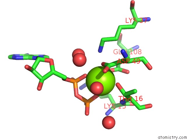
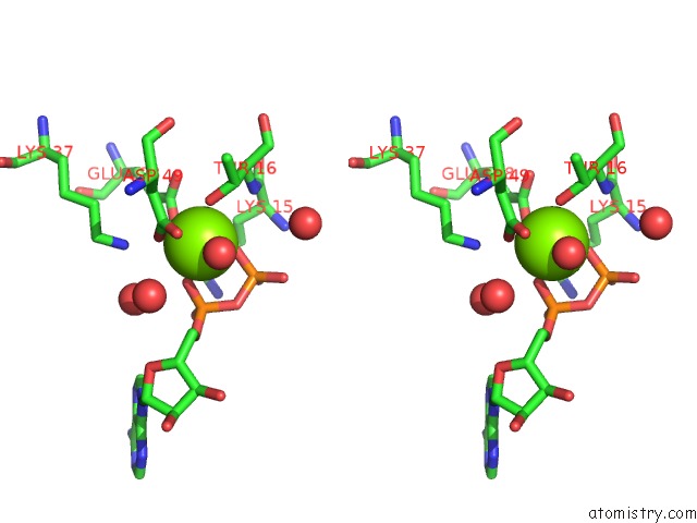
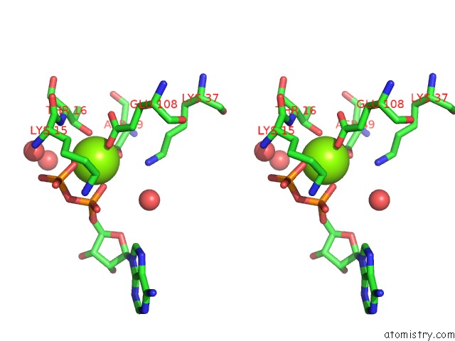
Magnesium binding site 1 out of 4 in 6czd
Go back to
Magnesium binding site 1 out
of 4 in the Crystal Structure of Mycobacterium Tuberculosis Dethiobiotin Synthetase in Complex with Adenosine Diphosphate

Mono view

Stereo pair view
Mono view
Stereo pair view
A full contact list of Magnesium with other atoms in the Mg binding
site number 1 of Crystal Structure of Mycobacterium Tuberculosis Dethiobiotin Synthetase in Complex with Adenosine Diphosphate within 5.0Å range:
|
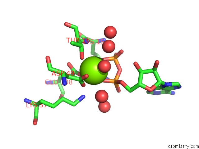
Magnesium binding site 2 out of 4 in 6czd
Go back to
Magnesium binding site 2 out
of 4 in the Crystal Structure of Mycobacterium Tuberculosis Dethiobiotin Synthetase in Complex with Adenosine Diphosphate
Mono view
Stereo pair view
Mono view
Stereo pair view
A full contact list of Magnesium with other atoms in the Mg binding
site number 2 of Crystal Structure of Mycobacterium Tuberculosis Dethiobiotin Synthetase in Complex with Adenosine Diphosphate within 5.0Å range:
|
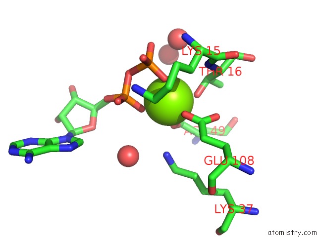
Magnesium binding site 3 out of 4 in 6czd
Go back to
Magnesium binding site 3 out
of 4 in the Crystal Structure of Mycobacterium Tuberculosis Dethiobiotin Synthetase in Complex with Adenosine Diphosphate
Mono view

Stereo pair view
Mono view
Stereo pair view
A full contact list of Magnesium with other atoms in the Mg binding
site number 3 of Crystal Structure of Mycobacterium Tuberculosis Dethiobiotin Synthetase in Complex with Adenosine Diphosphate within 5.0Å range:
|
Magnesium binding site 4 out of 4 in 6czd
Go back to
Magnesium binding site 4 out
of 4 in the Crystal Structure of Mycobacterium Tuberculosis Dethiobiotin Synthetase in Complex with Adenosine Diphosphate
Mono view
Stereo pair view
Mono view
Stereo pair view
A full contact list of Magnesium with other atoms in the Mg binding
site number 4 of Crystal Structure of Mycobacterium Tuberculosis Dethiobiotin Synthetase in Complex with Adenosine Diphosphate within 5.0Å range:
|
Reference:
A.P.Thompson,
K.L.Wegener,
G.W.Booker,
S.W.Polyak,
J.B.Bruning.
Precipitant-Ligand Exchange Technique Reveals the Adp Binding Mode in Mycobacterium Tuberculosis Dethiobiotin Synthetase. Acta Crystallogr D Struct V. 74 965 2018BIOL.
ISSN: ISSN 2059-7983
PubMed: 30289406
DOI: 10.1107/S2059798318010136
Page generated: Mon Sep 30 22:50:39 2024
ISSN: ISSN 2059-7983
PubMed: 30289406
DOI: 10.1107/S2059798318010136
Last articles
Zn in 9MJ5Zn in 9HNW
Zn in 9G0L
Zn in 9FNE
Zn in 9DZN
Zn in 9E0I
Zn in 9D32
Zn in 9DAK
Zn in 8ZXC
Zn in 8ZUF