Magnesium »
PDB 6d2y-6d9l »
6d3k »
Magnesium in PDB 6d3k: Crystal Structure of Unphosphorylated Human Pkr Kinase Domain in Complex with Adp
Enzymatic activity of Crystal Structure of Unphosphorylated Human Pkr Kinase Domain in Complex with Adp
All present enzymatic activity of Crystal Structure of Unphosphorylated Human Pkr Kinase Domain in Complex with Adp:
2.7.10.2; 2.7.11.1;
2.7.10.2; 2.7.11.1;
Protein crystallography data
The structure of Crystal Structure of Unphosphorylated Human Pkr Kinase Domain in Complex with Adp, PDB code: 6d3k
was solved by
H.Erlandsen,
C.B.Mayo,
V.L.Robinson,
J.L.Cole,
with X-Ray Crystallography technique. A brief refinement statistics is given in the table below:
| Resolution Low / High (Å) | 172.99 / 2.60 |
| Space group | C 2 2 21 |
| Cell size a, b, c (Å), α, β, γ (°) | 106.480, 159.600, 172.990, 90.00, 90.00, 90.00 |
| R / Rfree (%) | 20.9 / 26.6 |
Magnesium Binding Sites:
The binding sites of Magnesium atom in the Crystal Structure of Unphosphorylated Human Pkr Kinase Domain in Complex with Adp
(pdb code 6d3k). This binding sites where shown within
5.0 Angstroms radius around Magnesium atom.
In total 3 binding sites of Magnesium where determined in the Crystal Structure of Unphosphorylated Human Pkr Kinase Domain in Complex with Adp, PDB code: 6d3k:
Jump to Magnesium binding site number: 1; 2; 3;
In total 3 binding sites of Magnesium where determined in the Crystal Structure of Unphosphorylated Human Pkr Kinase Domain in Complex with Adp, PDB code: 6d3k:
Jump to Magnesium binding site number: 1; 2; 3;
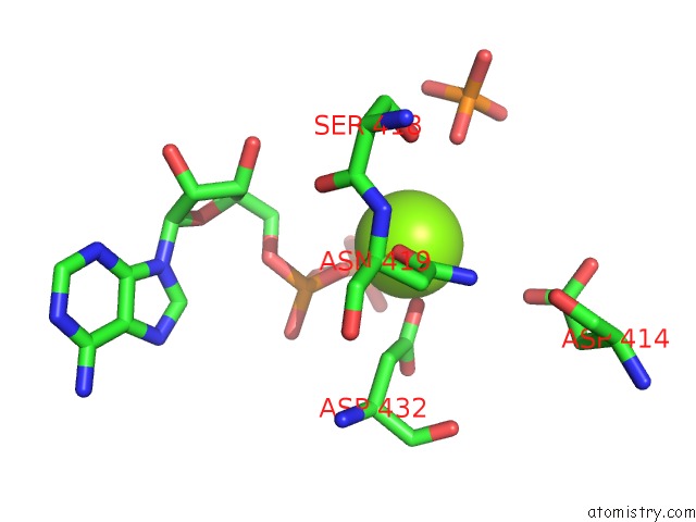
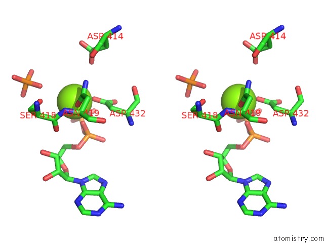
Magnesium binding site 1 out of 3 in 6d3k
Go back to
Magnesium binding site 1 out
of 3 in the Crystal Structure of Unphosphorylated Human Pkr Kinase Domain in Complex with Adp
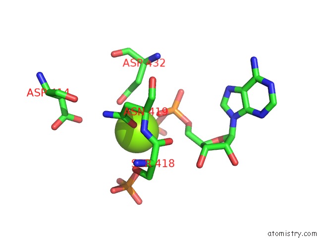
Mono view
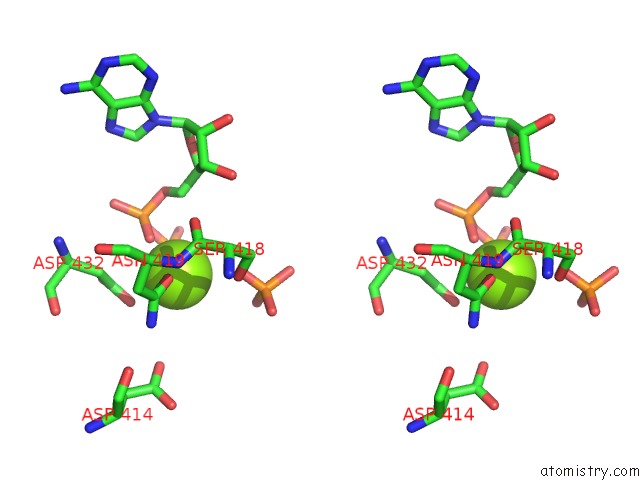
Stereo pair view
Mono view
Stereo pair view
A full contact list of Magnesium with other atoms in the Mg binding
site number 1 of Crystal Structure of Unphosphorylated Human Pkr Kinase Domain in Complex with Adp within 5.0Å range:
|
Magnesium binding site 2 out of 3 in 6d3k
Go back to
Magnesium binding site 2 out
of 3 in the Crystal Structure of Unphosphorylated Human Pkr Kinase Domain in Complex with Adp
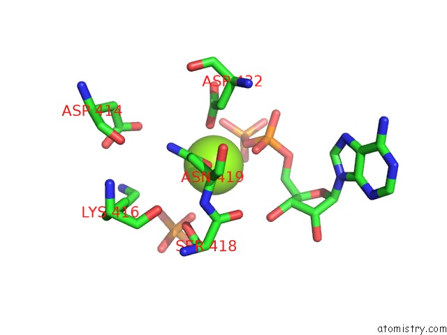
Mono view
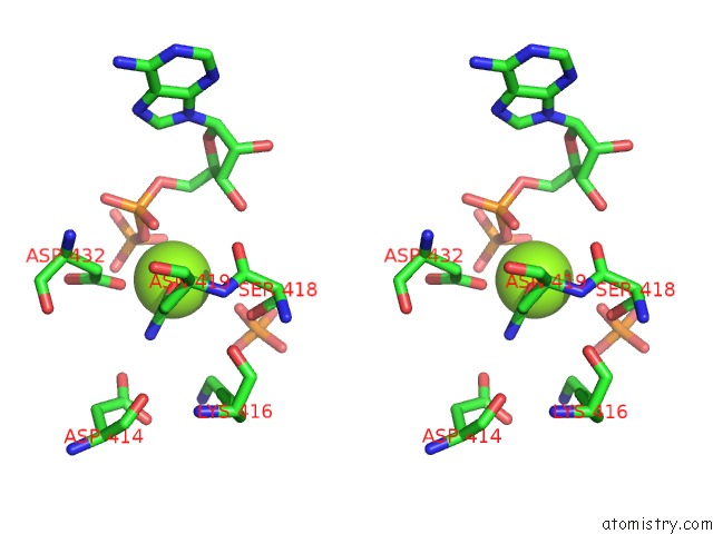
Stereo pair view
Mono view
Stereo pair view
A full contact list of Magnesium with other atoms in the Mg binding
site number 2 of Crystal Structure of Unphosphorylated Human Pkr Kinase Domain in Complex with Adp within 5.0Å range:
|
Magnesium binding site 3 out of 3 in 6d3k
Go back to
Magnesium binding site 3 out
of 3 in the Crystal Structure of Unphosphorylated Human Pkr Kinase Domain in Complex with Adp
Mono view
Stereo pair view
Mono view
Stereo pair view
A full contact list of Magnesium with other atoms in the Mg binding
site number 3 of Crystal Structure of Unphosphorylated Human Pkr Kinase Domain in Complex with Adp within 5.0Å range:
|
Reference:
C.B.Mayo,
H.Erlandsen,
D.J.Mouser,
A.G.Feinstein,
V.L.Robinson,
E.R.May,
J.L.Cole.
Structural Basis of Protein Kinase R Autophosphorylation. Biochemistry V. 58 2967 2019.
ISSN: ISSN 0006-2960
PubMed: 31246429
DOI: 10.1021/ACS.BIOCHEM.9B00161
Page generated: Mon Sep 30 22:53:11 2024
ISSN: ISSN 0006-2960
PubMed: 31246429
DOI: 10.1021/ACS.BIOCHEM.9B00161
Last articles
Zn in 9J0NZn in 9J0O
Zn in 9J0P
Zn in 9FJX
Zn in 9EKB
Zn in 9C0F
Zn in 9CAH
Zn in 9CH0
Zn in 9CH3
Zn in 9CH1