Magnesium »
PDB 6dw7-6e8s »
6dwt »
Magnesium in PDB 6dwt: Crystal Structure of Double-Stranded Dna Gaggcctc, Crystals Grown in MG2+
Protein crystallography data
The structure of Crystal Structure of Double-Stranded Dna Gaggcctc, Crystals Grown in MG2+, PDB code: 6dwt
was solved by
C.Hou,
O.V.Tsodikov,
with X-Ray Crystallography technique. A brief refinement statistics is given in the table below:
| Resolution Low / High (Å) | 30.00 / 1.60 |
| Space group | P 43 21 2 |
| Cell size a, b, c (Å), α, β, γ (°) | 44.255, 44.255, 23.597, 90.00, 90.00, 90.00 |
| R / Rfree (%) | 18.7 / 21.9 |
Magnesium Binding Sites:
The binding sites of Magnesium atom in the Crystal Structure of Double-Stranded Dna Gaggcctc, Crystals Grown in MG2+
(pdb code 6dwt). This binding sites where shown within
5.0 Angstroms radius around Magnesium atom.
In total only one binding site of Magnesium was determined in the Crystal Structure of Double-Stranded Dna Gaggcctc, Crystals Grown in MG2+, PDB code: 6dwt:
In total only one binding site of Magnesium was determined in the Crystal Structure of Double-Stranded Dna Gaggcctc, Crystals Grown in MG2+, PDB code: 6dwt:
Magnesium binding site 1 out of 1 in 6dwt
Go back to
Magnesium binding site 1 out
of 1 in the Crystal Structure of Double-Stranded Dna Gaggcctc, Crystals Grown in MG2+
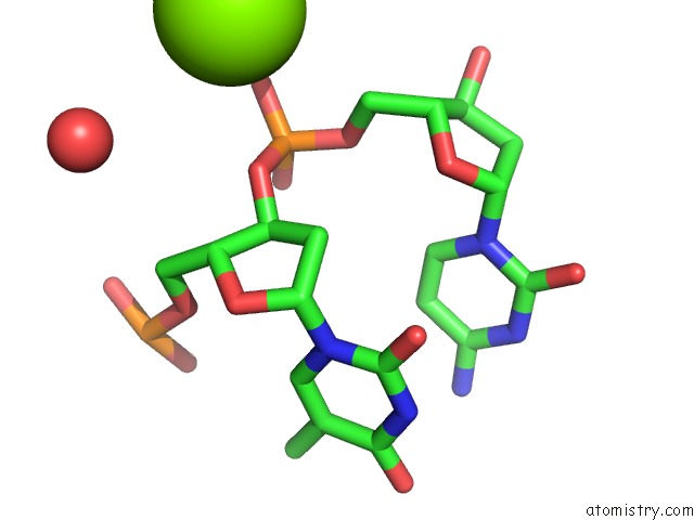
Mono view
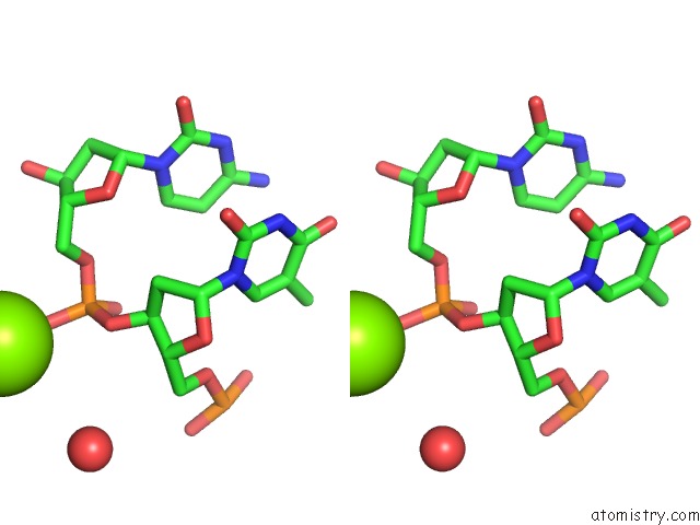
Stereo pair view
Mono view
Stereo pair view
A full contact list of Magnesium with other atoms in the Mg binding
site number 1 of Crystal Structure of Double-Stranded Dna Gaggcctc, Crystals Grown in MG2+ within 5.0Å range:
|
Reference:
C.Hou,
O.V.Tsodikov.
Utilizing Guanine Coordinated ZN2+ to Determine Dna Crystal Structures By Single-Wavelength Anomalous Diffraction Acta Crystallogr.,Sect.D V. 75 32 2019.
ISSN: ISSN 0907-4449
Page generated: Mon Sep 30 23:42:56 2024
ISSN: ISSN 0907-4449
Last articles
Zn in 9MJ5Zn in 9HNW
Zn in 9G0L
Zn in 9FNE
Zn in 9DZN
Zn in 9E0I
Zn in 9D32
Zn in 9DAK
Zn in 8ZXC
Zn in 8ZUF