Magnesium »
PDB 6dw7-6e8s »
6e20 »
Magnesium in PDB 6e20: Crystal Structure of the Dario Rerio Galectin-1-L2
Protein crystallography data
The structure of Crystal Structure of the Dario Rerio Galectin-1-L2, PDB code: 6e20
was solved by
A.Ghosh,
M.A.Bianchet,
with X-Ray Crystallography technique. A brief refinement statistics is given in the table below:
| Resolution Low / High (Å) | 34.81 / 2.00 |
| Space group | P 61 |
| Cell size a, b, c (Å), α, β, γ (°) | 40.438, 40.438, 303.639, 90.00, 90.00, 120.00 |
| R / Rfree (%) | 17.4 / 23.4 |
Magnesium Binding Sites:
The binding sites of Magnesium atom in the Crystal Structure of the Dario Rerio Galectin-1-L2
(pdb code 6e20). This binding sites where shown within
5.0 Angstroms radius around Magnesium atom.
In total 2 binding sites of Magnesium where determined in the Crystal Structure of the Dario Rerio Galectin-1-L2, PDB code: 6e20:
Jump to Magnesium binding site number: 1; 2;
In total 2 binding sites of Magnesium where determined in the Crystal Structure of the Dario Rerio Galectin-1-L2, PDB code: 6e20:
Jump to Magnesium binding site number: 1; 2;
Magnesium binding site 1 out of 2 in 6e20
Go back to
Magnesium binding site 1 out
of 2 in the Crystal Structure of the Dario Rerio Galectin-1-L2
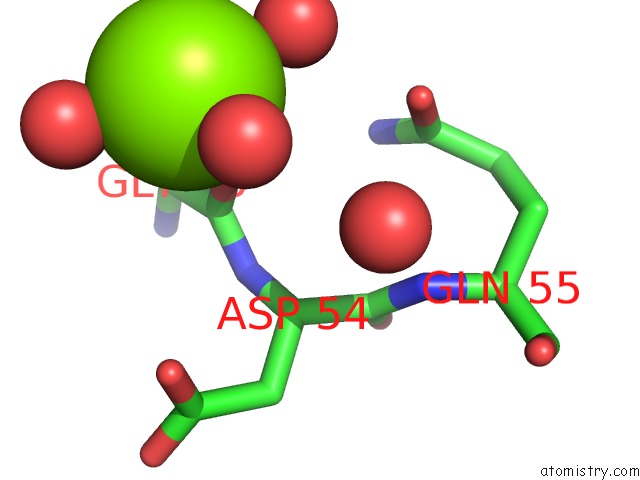
Mono view
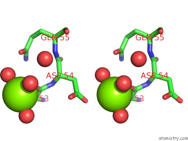
Stereo pair view
Mono view
Stereo pair view
A full contact list of Magnesium with other atoms in the Mg binding
site number 1 of Crystal Structure of the Dario Rerio Galectin-1-L2 within 5.0Å range:
|
Magnesium binding site 2 out of 2 in 6e20
Go back to
Magnesium binding site 2 out
of 2 in the Crystal Structure of the Dario Rerio Galectin-1-L2
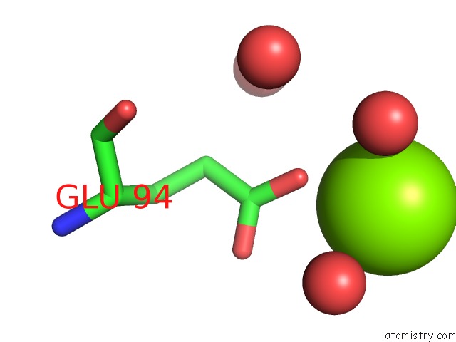
Mono view
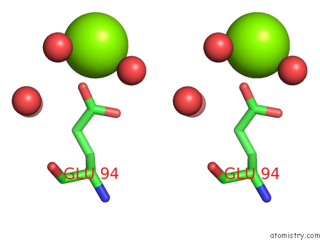
Stereo pair view
Mono view
Stereo pair view
A full contact list of Magnesium with other atoms in the Mg binding
site number 2 of Crystal Structure of the Dario Rerio Galectin-1-L2 within 5.0Å range:
|
Reference:
A.Ghosh,
A.Banerjee,
L.M.Amzel,
G.R.Vasta,
M.A.Bianchet.
Structure of the Zebrafish Galectin-1-L2 and Model of Its Interaction with the Infectious Hematopoietic Necrosis Virus (Ihnv) Envelope Glycoprotein. Glycobiology V. 29 419 2019.
ISSN: ESSN 1460-2423
PubMed: 30834446
DOI: 10.1093/GLYCOB/CWZ015
Page generated: Mon Sep 30 23:47:45 2024
ISSN: ESSN 1460-2423
PubMed: 30834446
DOI: 10.1093/GLYCOB/CWZ015
Last articles
Zn in 9MJ5Zn in 9HNW
Zn in 9G0L
Zn in 9FNE
Zn in 9DZN
Zn in 9E0I
Zn in 9D32
Zn in 9DAK
Zn in 8ZXC
Zn in 8ZUF