Magnesium »
PDB 6f6u-6fch »
6f7t »
Magnesium in PDB 6f7t: Crystal Structure of An Fab Fragment in Complex with A Peptide From Bacillus Subtilis Rnase Y
Protein crystallography data
The structure of Crystal Structure of An Fab Fragment in Complex with A Peptide From Bacillus Subtilis Rnase Y, PDB code: 6f7t
was solved by
B.Golinelli-Pimpaneau,
P.Hardouin,
with X-Ray Crystallography technique. A brief refinement statistics is given in the table below:
| Resolution Low / High (Å) | 46.15 / 2.60 |
| Space group | P 21 21 2 |
| Cell size a, b, c (Å), α, β, γ (°) | 98.680, 130.450, 72.050, 90.00, 90.00, 90.00 |
| R / Rfree (%) | 20.2 / 25.3 |
Magnesium Binding Sites:
The binding sites of Magnesium atom in the Crystal Structure of An Fab Fragment in Complex with A Peptide From Bacillus Subtilis Rnase Y
(pdb code 6f7t). This binding sites where shown within
5.0 Angstroms radius around Magnesium atom.
In total only one binding site of Magnesium was determined in the Crystal Structure of An Fab Fragment in Complex with A Peptide From Bacillus Subtilis Rnase Y, PDB code: 6f7t:
In total only one binding site of Magnesium was determined in the Crystal Structure of An Fab Fragment in Complex with A Peptide From Bacillus Subtilis Rnase Y, PDB code: 6f7t:
Magnesium binding site 1 out of 1 in 6f7t
Go back to
Magnesium binding site 1 out
of 1 in the Crystal Structure of An Fab Fragment in Complex with A Peptide From Bacillus Subtilis Rnase Y
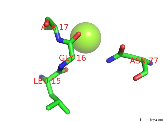
Mono view
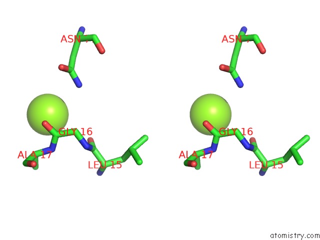
Stereo pair view
Mono view
Stereo pair view
A full contact list of Magnesium with other atoms in the Mg binding
site number 1 of Crystal Structure of An Fab Fragment in Complex with A Peptide From Bacillus Subtilis Rnase Y within 5.0Å range:
|
Reference:
P.Hardouin,
C.Velours,
C.Bou-Nader,
N.Assrir,
S.Laalami,
H.Putzer,
D.Durand,
B.Golinelli-Pimpaneau.
Dissociation of the Dimer of the Intrinsically Disordered Domain of Rnase Y Upon Antibody Binding. Biophys. J. V. 115 2102 2018.
ISSN: ESSN 1542-0086
PubMed: 30447990
DOI: 10.1016/J.BPJ.2018.10.016
Page generated: Tue Oct 1 00:10:53 2024
ISSN: ESSN 1542-0086
PubMed: 30447990
DOI: 10.1016/J.BPJ.2018.10.016
Last articles
Ca in 5UBOCa in 5U95
Ca in 5UBA
Ca in 5UB5
Ca in 5U8O
Ca in 5U6J
Ca in 5U5N
Ca in 5U5I
Ca in 5U3K
Ca in 5U3E