Magnesium »
PDB 6f6u-6fch »
6f8v »
Magnesium in PDB 6f8v: Crystal Structure of the PDE4D Catalytic Domain in Complex with Gebr- 18B
Enzymatic activity of Crystal Structure of the PDE4D Catalytic Domain in Complex with Gebr- 18B
All present enzymatic activity of Crystal Structure of the PDE4D Catalytic Domain in Complex with Gebr- 18B:
3.1.4.53;
3.1.4.53;
Protein crystallography data
The structure of Crystal Structure of the PDE4D Catalytic Domain in Complex with Gebr- 18B, PDB code: 6f8v
was solved by
T.Prosdocimi,
S.Donini,
E.Parisini,
with X-Ray Crystallography technique. A brief refinement statistics is given in the table below:
| Resolution Low / High (Å) | 45.49 / 1.85 |
| Space group | P 21 21 21 |
| Cell size a, b, c (Å), α, β, γ (°) | 64.558, 98.314, 120.089, 90.00, 90.00, 90.00 |
| R / Rfree (%) | 20.5 / 25.4 |
Other elements in 6f8v:
The structure of Crystal Structure of the PDE4D Catalytic Domain in Complex with Gebr- 18B also contains other interesting chemical elements:
| Zinc | (Zn) | 2 atoms |
Magnesium Binding Sites:
The binding sites of Magnesium atom in the Crystal Structure of the PDE4D Catalytic Domain in Complex with Gebr- 18B
(pdb code 6f8v). This binding sites where shown within
5.0 Angstroms radius around Magnesium atom.
In total 4 binding sites of Magnesium where determined in the Crystal Structure of the PDE4D Catalytic Domain in Complex with Gebr- 18B, PDB code: 6f8v:
Jump to Magnesium binding site number: 1; 2; 3; 4;
In total 4 binding sites of Magnesium where determined in the Crystal Structure of the PDE4D Catalytic Domain in Complex with Gebr- 18B, PDB code: 6f8v:
Jump to Magnesium binding site number: 1; 2; 3; 4;
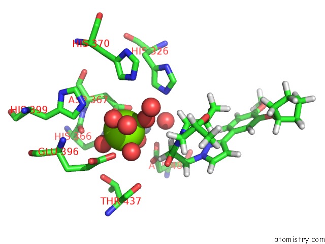
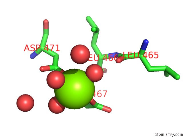
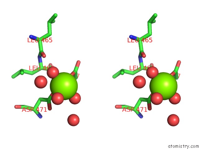
Magnesium binding site 1 out of 4 in 6f8v
Go back to
Magnesium binding site 1 out
of 4 in the Crystal Structure of the PDE4D Catalytic Domain in Complex with Gebr- 18B
Mono view
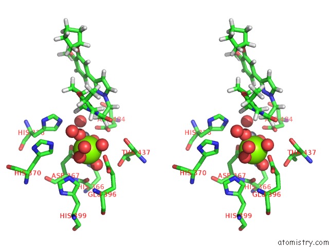
Stereo pair view
Mono view
Stereo pair view
A full contact list of Magnesium with other atoms in the Mg binding
site number 1 of Crystal Structure of the PDE4D Catalytic Domain in Complex with Gebr- 18B within 5.0Å range:
|
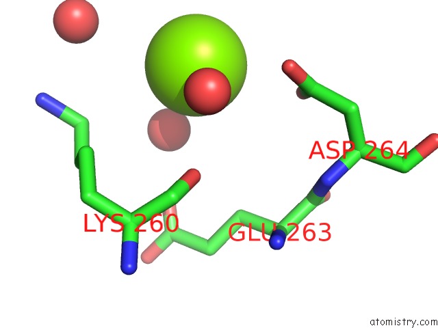
Magnesium binding site 2 out of 4 in 6f8v
Go back to
Magnesium binding site 2 out
of 4 in the Crystal Structure of the PDE4D Catalytic Domain in Complex with Gebr- 18B
Mono view
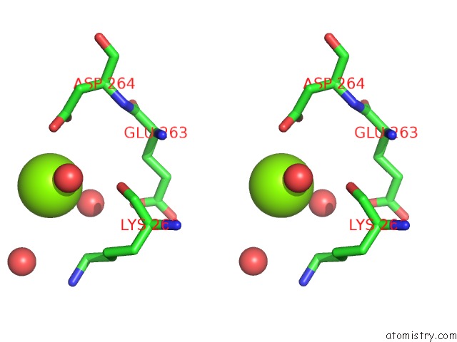
Stereo pair view
Mono view
Stereo pair view
A full contact list of Magnesium with other atoms in the Mg binding
site number 2 of Crystal Structure of the PDE4D Catalytic Domain in Complex with Gebr- 18B within 5.0Å range:
|
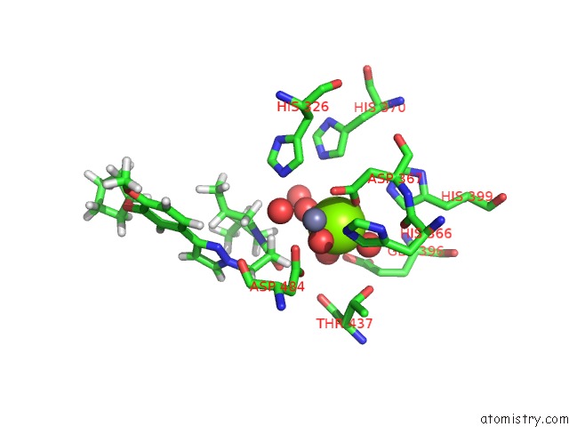
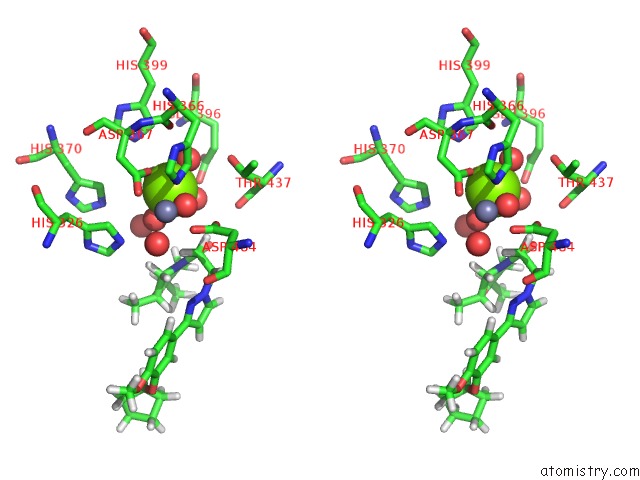
Magnesium binding site 3 out of 4 in 6f8v
Go back to
Magnesium binding site 3 out
of 4 in the Crystal Structure of the PDE4D Catalytic Domain in Complex with Gebr- 18B
Mono view
Stereo pair view
Mono view
Stereo pair view
A full contact list of Magnesium with other atoms in the Mg binding
site number 3 of Crystal Structure of the PDE4D Catalytic Domain in Complex with Gebr- 18B within 5.0Å range:
|
Magnesium binding site 4 out of 4 in 6f8v
Go back to
Magnesium binding site 4 out
of 4 in the Crystal Structure of the PDE4D Catalytic Domain in Complex with Gebr- 18B
Mono view
Stereo pair view
Mono view
Stereo pair view
A full contact list of Magnesium with other atoms in the Mg binding
site number 4 of Crystal Structure of the PDE4D Catalytic Domain in Complex with Gebr- 18B within 5.0Å range:
|
Reference:
T.Prosdocimi,
L.Mollica,
S.Donini,
M.S.Semrau,
A.P.Lucarelli,
E.Aiolfi,
A.Cavalli,
P.Storici,
S.Alfei,
C.Brullo,
O.Bruno,
E.Parisini.
Molecular Bases of PDE4D Inhibition By Memory-Enhancing Gebr Library Compounds. Biochemistry V. 57 2876 2018.
ISSN: ISSN 1520-4995
PubMed: 29652483
DOI: 10.1021/ACS.BIOCHEM.8B00288
Page generated: Tue Oct 1 00:12:00 2024
ISSN: ISSN 1520-4995
PubMed: 29652483
DOI: 10.1021/ACS.BIOCHEM.8B00288
Last articles
Cl in 5Y00Cl in 5XZI
Cl in 5XZG
Cl in 5XYZ
Cl in 5XZJ
Cl in 5XWM
Cl in 5XYY
Cl in 5XYG
Cl in 5XYX
Cl in 5XYR