Magnesium »
PDB 6fch-6fjk »
6fd4 »
Magnesium in PDB 6fd4: Crystal Structure of Human Aprt-TYR105PHE Variant in Complex with Adenine, Prpp and MG2+, 14 Hours Post Crystallization
Enzymatic activity of Crystal Structure of Human Aprt-TYR105PHE Variant in Complex with Adenine, Prpp and MG2+, 14 Hours Post Crystallization
All present enzymatic activity of Crystal Structure of Human Aprt-TYR105PHE Variant in Complex with Adenine, Prpp and MG2+, 14 Hours Post Crystallization:
2.4.2.7;
2.4.2.7;
Protein crystallography data
The structure of Crystal Structure of Human Aprt-TYR105PHE Variant in Complex with Adenine, Prpp and MG2+, 14 Hours Post Crystallization, PDB code: 6fd4
was solved by
P.Nioche,
J.Huyet,
M.Ozeir,
with X-Ray Crystallography technique. A brief refinement statistics is given in the table below:
| Resolution Low / High (Å) | 44.64 / 1.50 |
| Space group | P 1 |
| Cell size a, b, c (Å), α, β, γ (°) | 47.380, 47.540, 47.670, 69.86, 77.20, 61.76 |
| R / Rfree (%) | 16.6 / 17.8 |
Magnesium Binding Sites:
The binding sites of Magnesium atom in the Crystal Structure of Human Aprt-TYR105PHE Variant in Complex with Adenine, Prpp and MG2+, 14 Hours Post Crystallization
(pdb code 6fd4). This binding sites where shown within
5.0 Angstroms radius around Magnesium atom.
In total 2 binding sites of Magnesium where determined in the Crystal Structure of Human Aprt-TYR105PHE Variant in Complex with Adenine, Prpp and MG2+, 14 Hours Post Crystallization, PDB code: 6fd4:
Jump to Magnesium binding site number: 1; 2;
In total 2 binding sites of Magnesium where determined in the Crystal Structure of Human Aprt-TYR105PHE Variant in Complex with Adenine, Prpp and MG2+, 14 Hours Post Crystallization, PDB code: 6fd4:
Jump to Magnesium binding site number: 1; 2;
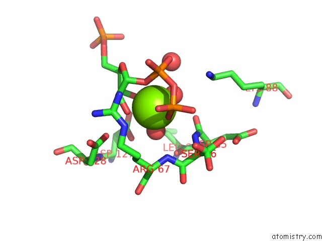
Magnesium binding site 1 out of 2 in 6fd4
Go back to
Magnesium binding site 1 out
of 2 in the Crystal Structure of Human Aprt-TYR105PHE Variant in Complex with Adenine, Prpp and MG2+, 14 Hours Post Crystallization
Mono view

Stereo pair view
Mono view
Stereo pair view
A full contact list of Magnesium with other atoms in the Mg binding
site number 1 of Crystal Structure of Human Aprt-TYR105PHE Variant in Complex with Adenine, Prpp and MG2+, 14 Hours Post Crystallization within 5.0Å range:
|
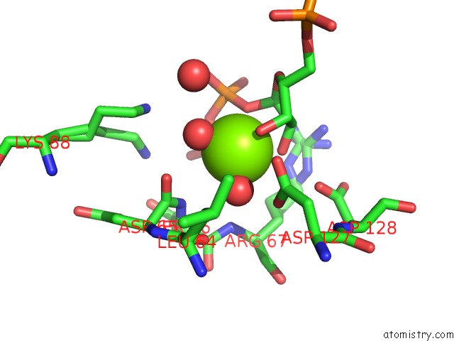
Magnesium binding site 2 out of 2 in 6fd4
Go back to
Magnesium binding site 2 out
of 2 in the Crystal Structure of Human Aprt-TYR105PHE Variant in Complex with Adenine, Prpp and MG2+, 14 Hours Post Crystallization
Mono view

Stereo pair view
Mono view
Stereo pair view
A full contact list of Magnesium with other atoms in the Mg binding
site number 2 of Crystal Structure of Human Aprt-TYR105PHE Variant in Complex with Adenine, Prpp and MG2+, 14 Hours Post Crystallization within 5.0Å range:
|
Reference:
J.Huyet,
M.Ozeir,
M.C.Burgevin,
B.Pinson,
F.Chesney,
J.M.Remy,
A.R.Siddiqi,
R.Lupoli,
G.Pinon,
C.Saint-Marc,
J.F.Gibert,
R.Morales,
I.Ceballos-Picot,
R.Barouki,
B.Daignan-Fornier,
A.Olivier-Bandini,
F.Auge,
P.Nioche.
Structural Insights Into the Forward and Reverse Enzymatic Reactions in Human Adenine Phosphoribosyltransferase. Cell Chem Biol V. 25 666 2018.
ISSN: ESSN 2451-9448
PubMed: 29576532
DOI: 10.1016/J.CHEMBIOL.2018.02.011
Page generated: Tue Oct 1 00:23:54 2024
ISSN: ESSN 2451-9448
PubMed: 29576532
DOI: 10.1016/J.CHEMBIOL.2018.02.011
Last articles
Zn in 9J0NZn in 9J0O
Zn in 9J0P
Zn in 9FJX
Zn in 9EKB
Zn in 9C0F
Zn in 9CAH
Zn in 9CH0
Zn in 9CH3
Zn in 9CH1