Magnesium »
PDB 6fjm-6ftm »
6fk5 »
Magnesium in PDB 6fk5: Structure of 3' Phosphatase Nexo (D146N) From Neisseria Bound to Dna Substrate in Presence of Magnesium Ion
Protein crystallography data
The structure of Structure of 3' Phosphatase Nexo (D146N) From Neisseria Bound to Dna Substrate in Presence of Magnesium Ion, PDB code: 6fk5
was solved by
J.Silhan,
Q.Zhao,
E.Boura,
H.Thomson,
A.Foster,
C.M.Tang,
P.S.Freemont,
G.S.Baldwin,
with X-Ray Crystallography technique. A brief refinement statistics is given in the table below:
| Resolution Low / High (Å) | 50.43 / 2.02 |
| Space group | P 43 21 2 |
| Cell size a, b, c (Å), α, β, γ (°) | 58.940, 58.940, 292.260, 90.00, 90.00, 90.00 |
| R / Rfree (%) | 23 / 25.8 |
Magnesium Binding Sites:
The binding sites of Magnesium atom in the Structure of 3' Phosphatase Nexo (D146N) From Neisseria Bound to Dna Substrate in Presence of Magnesium Ion
(pdb code 6fk5). This binding sites where shown within
5.0 Angstroms radius around Magnesium atom.
In total 2 binding sites of Magnesium where determined in the Structure of 3' Phosphatase Nexo (D146N) From Neisseria Bound to Dna Substrate in Presence of Magnesium Ion, PDB code: 6fk5:
Jump to Magnesium binding site number: 1; 2;
In total 2 binding sites of Magnesium where determined in the Structure of 3' Phosphatase Nexo (D146N) From Neisseria Bound to Dna Substrate in Presence of Magnesium Ion, PDB code: 6fk5:
Jump to Magnesium binding site number: 1; 2;
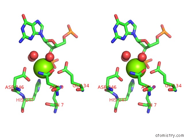
Magnesium binding site 1 out of 2 in 6fk5
Go back to
Magnesium binding site 1 out
of 2 in the Structure of 3' Phosphatase Nexo (D146N) From Neisseria Bound to Dna Substrate in Presence of Magnesium Ion

Mono view
Stereo pair view
Mono view
Stereo pair view
A full contact list of Magnesium with other atoms in the Mg binding
site number 1 of Structure of 3' Phosphatase Nexo (D146N) From Neisseria Bound to Dna Substrate in Presence of Magnesium Ion within 5.0Å range:
|
Magnesium binding site 2 out of 2 in 6fk5
Go back to
Magnesium binding site 2 out
of 2 in the Structure of 3' Phosphatase Nexo (D146N) From Neisseria Bound to Dna Substrate in Presence of Magnesium Ion

Mono view

Stereo pair view
Mono view
Stereo pair view
A full contact list of Magnesium with other atoms in the Mg binding
site number 2 of Structure of 3' Phosphatase Nexo (D146N) From Neisseria Bound to Dna Substrate in Presence of Magnesium Ion within 5.0Å range:
|
Reference:
J.Silhan,
Q.Zhao,
E.Boura,
H.Thomson,
A.Forster,
C.M.Tang,
P.S.Freemont,
G.S.Baldwin.
Structural Basis For Recognition and Repair of the 3'-Phosphate By Nexo, A Base Excision Dna Repair Nuclease From Neisseria Meningitidis. Nucleic Acids Res. V. 46 11980 2018.
ISSN: ESSN 1362-4962
PubMed: 30329088
DOI: 10.1093/NAR/GKY934
Page generated: Tue Oct 1 00:31:19 2024
ISSN: ESSN 1362-4962
PubMed: 30329088
DOI: 10.1093/NAR/GKY934
Last articles
Zn in 9MJ5Zn in 9HNW
Zn in 9G0L
Zn in 9FNE
Zn in 9DZN
Zn in 9E0I
Zn in 9D32
Zn in 9DAK
Zn in 8ZXC
Zn in 8ZUF