Magnesium »
PDB 6ftm-6g6y »
6fwz »
Magnesium in PDB 6fwz: Crystal Structure of Human Udp-N-Acetylglucosamine-Dolichyl-Phosphate N-Acetylglucosaminephosphotransferase (DPAGT1) (V264G Mutant) in Complex with Udp-Glcnac
Enzymatic activity of Crystal Structure of Human Udp-N-Acetylglucosamine-Dolichyl-Phosphate N-Acetylglucosaminephosphotransferase (DPAGT1) (V264G Mutant) in Complex with Udp-Glcnac
All present enzymatic activity of Crystal Structure of Human Udp-N-Acetylglucosamine-Dolichyl-Phosphate N-Acetylglucosaminephosphotransferase (DPAGT1) (V264G Mutant) in Complex with Udp-Glcnac:
2.7.8.15;
2.7.8.15;
Protein crystallography data
The structure of Crystal Structure of Human Udp-N-Acetylglucosamine-Dolichyl-Phosphate N-Acetylglucosaminephosphotransferase (DPAGT1) (V264G Mutant) in Complex with Udp-Glcnac, PDB code: 6fwz
was solved by
A.C.W.Pike,
Y.Y.Dong,
A.Chu,
A.Tessitore,
S.Goubin,
L.Dong,
S.Mukhopadhyay,
P.Mahajan,
R.Chalk,
G.Berridge,
D.Wang,
K.Kupinska,
K.Belaya,
D.Beeson,
N.Burgess-Brown,
A.M.Edwards,
C.H.Arrowsmith,
C.Bountra,
E.P.Carpenter,
Structural Genomics Consortium (Sgc),
with X-Ray Crystallography technique. A brief refinement statistics is given in the table below:
| Resolution Low / High (Å) | 30.00 / 3.10 |
| Space group | P 65 2 2 |
| Cell size a, b, c (Å), α, β, γ (°) | 102.459, 102.459, 238.209, 90.00, 90.00, 120.00 |
| R / Rfree (%) | 22.3 / 23.6 |
Magnesium Binding Sites:
The binding sites of Magnesium atom in the Crystal Structure of Human Udp-N-Acetylglucosamine-Dolichyl-Phosphate N-Acetylglucosaminephosphotransferase (DPAGT1) (V264G Mutant) in Complex with Udp-Glcnac
(pdb code 6fwz). This binding sites where shown within
5.0 Angstroms radius around Magnesium atom.
In total only one binding site of Magnesium was determined in the Crystal Structure of Human Udp-N-Acetylglucosamine-Dolichyl-Phosphate N-Acetylglucosaminephosphotransferase (DPAGT1) (V264G Mutant) in Complex with Udp-Glcnac, PDB code: 6fwz:
In total only one binding site of Magnesium was determined in the Crystal Structure of Human Udp-N-Acetylglucosamine-Dolichyl-Phosphate N-Acetylglucosaminephosphotransferase (DPAGT1) (V264G Mutant) in Complex with Udp-Glcnac, PDB code: 6fwz:
Magnesium binding site 1 out of 1 in 6fwz
Go back to
Magnesium binding site 1 out
of 1 in the Crystal Structure of Human Udp-N-Acetylglucosamine-Dolichyl-Phosphate N-Acetylglucosaminephosphotransferase (DPAGT1) (V264G Mutant) in Complex with Udp-Glcnac
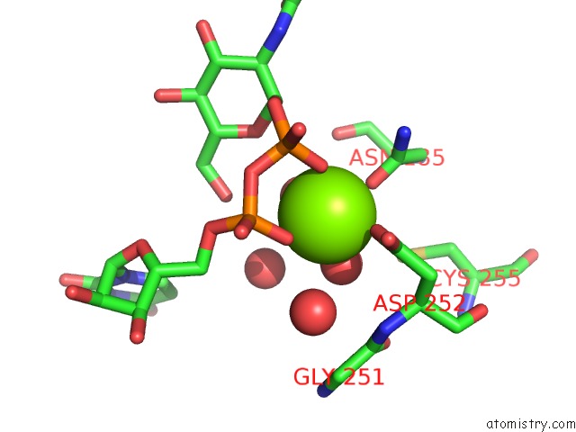
Mono view
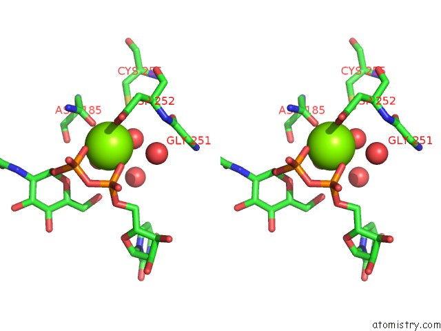
Stereo pair view
Mono view
Stereo pair view
A full contact list of Magnesium with other atoms in the Mg binding
site number 1 of Crystal Structure of Human Udp-N-Acetylglucosamine-Dolichyl-Phosphate N-Acetylglucosaminephosphotransferase (DPAGT1) (V264G Mutant) in Complex with Udp-Glcnac within 5.0Å range:
|
Reference:
Y.Y.Dong,
H.Wang,
A.C.W.Pike,
S.A.Cochrane,
S.Hamedzadeh,
F.J.Wyszynski,
S.R.Bushell,
S.F.Royer,
D.A.Widdick,
A.Sajid,
H.I.Boshoff,
Y.Park,
R.Lucas,
W.M.Liu,
S.S.Lee,
T.Machida,
L.Minall,
S.Mehmood,
K.Belaya,
W.W.Liu,
A.Chu,
L.Shrestha,
S.M.M.Mukhopadhyay,
C.Strain-Damerell,
R.Chalk,
N.A.Burgess-Brown,
M.J.Bibb,
C.E.Barry Iii,
C.V.Robinson,
D.Beeson,
B.G.Davis,
E.P.Carpenter.
Structures of DPAGT1 Explain Glycosylation Disease Mechanisms and Advance Tb Antibiotic Design. Cell V. 175 1045 2018.
ISSN: ISSN 1097-4172
PubMed: 30388443
DOI: 10.1016/J.CELL.2018.10.037
Page generated: Tue Oct 1 00:54:41 2024
ISSN: ISSN 1097-4172
PubMed: 30388443
DOI: 10.1016/J.CELL.2018.10.037
Last articles
Zn in 9J0NZn in 9J0O
Zn in 9J0P
Zn in 9FJX
Zn in 9EKB
Zn in 9C0F
Zn in 9CAH
Zn in 9CH0
Zn in 9CH3
Zn in 9CH1