Magnesium »
PDB 6ftm-6g6y »
6g2n »
Magnesium in PDB 6g2n: Crystal Structure of Human Cytosolic 5'(3')-Deoxyribonucleotidase in Complex with the Inhibitor Pb-Pau
Protein crystallography data
The structure of Crystal Structure of Human Cytosolic 5'(3')-Deoxyribonucleotidase in Complex with the Inhibitor Pb-Pau, PDB code: 6g2n
was solved by
P.Pachl,
P.Rezacova,
J.Brynda,
with X-Ray Crystallography technique. A brief refinement statistics is given in the table below:
| Resolution Low / High (Å) | 42.41 / 1.40 |
| Space group | P 1 |
| Cell size a, b, c (Å), α, β, γ (°) | 38.877, 47.231, 61.795, 67.86, 88.69, 77.37 |
| R / Rfree (%) | 15.1 / 19.2 |
Magnesium Binding Sites:
The binding sites of Magnesium atom in the Crystal Structure of Human Cytosolic 5'(3')-Deoxyribonucleotidase in Complex with the Inhibitor Pb-Pau
(pdb code 6g2n). This binding sites where shown within
5.0 Angstroms radius around Magnesium atom.
In total 2 binding sites of Magnesium where determined in the Crystal Structure of Human Cytosolic 5'(3')-Deoxyribonucleotidase in Complex with the Inhibitor Pb-Pau, PDB code: 6g2n:
Jump to Magnesium binding site number: 1; 2;
In total 2 binding sites of Magnesium where determined in the Crystal Structure of Human Cytosolic 5'(3')-Deoxyribonucleotidase in Complex with the Inhibitor Pb-Pau, PDB code: 6g2n:
Jump to Magnesium binding site number: 1; 2;
Magnesium binding site 1 out of 2 in 6g2n
Go back to
Magnesium binding site 1 out
of 2 in the Crystal Structure of Human Cytosolic 5'(3')-Deoxyribonucleotidase in Complex with the Inhibitor Pb-Pau
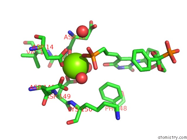
Mono view
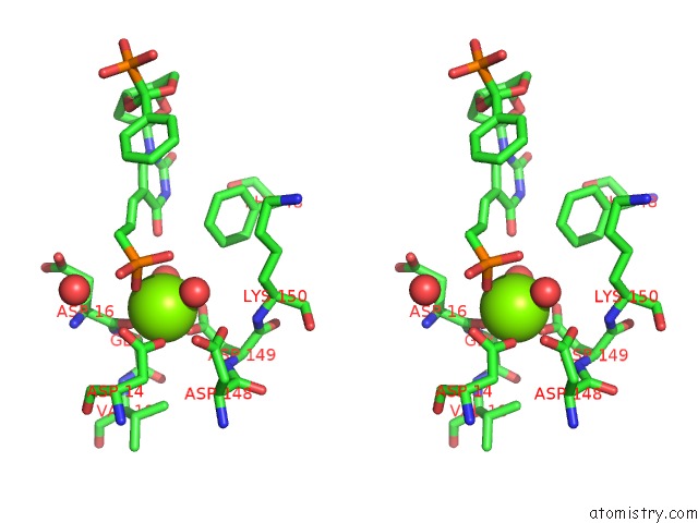
Stereo pair view
Mono view
Stereo pair view
A full contact list of Magnesium with other atoms in the Mg binding
site number 1 of Crystal Structure of Human Cytosolic 5'(3')-Deoxyribonucleotidase in Complex with the Inhibitor Pb-Pau within 5.0Å range:
|
Magnesium binding site 2 out of 2 in 6g2n
Go back to
Magnesium binding site 2 out
of 2 in the Crystal Structure of Human Cytosolic 5'(3')-Deoxyribonucleotidase in Complex with the Inhibitor Pb-Pau
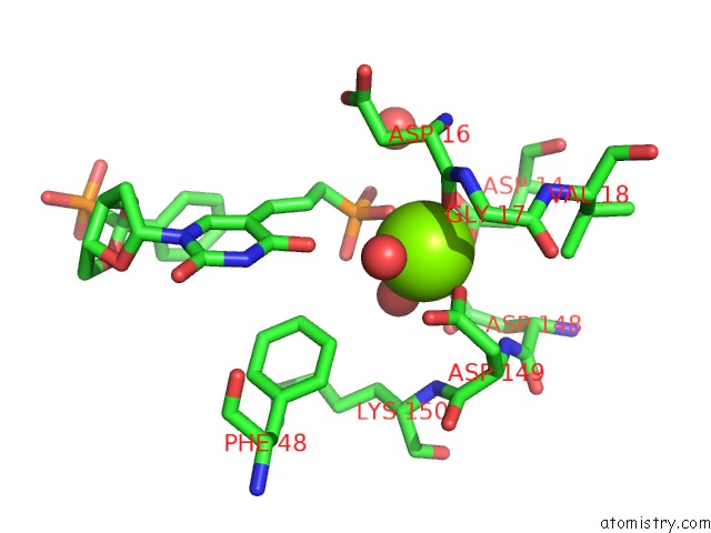
Mono view
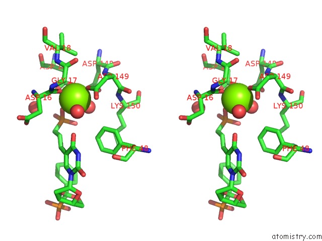
Stereo pair view
Mono view
Stereo pair view
A full contact list of Magnesium with other atoms in the Mg binding
site number 2 of Crystal Structure of Human Cytosolic 5'(3')-Deoxyribonucleotidase in Complex with the Inhibitor Pb-Pau within 5.0Å range:
|
Reference:
P.Pachl,
O.Simak,
M.Budesinsky,
J.Brynda,
I.Rosenberg,
P.Rezacova.
Structure-Based Optimization of Bisphosphonate Nucleoside Inhibitors of Human 5'(3')-Deoxyribonucleotidases Eur.J.Org.Chem. 2018.
ISSN: ISSN 1434-193X
DOI: 10.1002/EJOC.201800515
Page generated: Tue Oct 1 00:56:32 2024
ISSN: ISSN 1434-193X
DOI: 10.1002/EJOC.201800515
Last articles
Zn in 9J0NZn in 9J0O
Zn in 9J0P
Zn in 9FJX
Zn in 9EKB
Zn in 9C0F
Zn in 9CAH
Zn in 9CH0
Zn in 9CH3
Zn in 9CH1