Magnesium »
PDB 6g7d-6gg6 »
6g94 »
Magnesium in PDB 6g94: Structure of E. Coli Hydrogenase-1 C19G Variant in Complex with Cytochrome B
Enzymatic activity of Structure of E. Coli Hydrogenase-1 C19G Variant in Complex with Cytochrome B
All present enzymatic activity of Structure of E. Coli Hydrogenase-1 C19G Variant in Complex with Cytochrome B:
1.12.99.6;
1.12.99.6;
Protein crystallography data
The structure of Structure of E. Coli Hydrogenase-1 C19G Variant in Complex with Cytochrome B, PDB code: 6g94
was solved by
A.Volbeda,
J.C.Fontecilla-Camps,
with X-Ray Crystallography technique. A brief refinement statistics is given in the table below:
| Resolution Low / High (Å) | 25.00 / 2.50 |
| Space group | P 21 21 21 |
| Cell size a, b, c (Å), α, β, γ (°) | 124.410, 165.030, 206.220, 90.00, 90.00, 90.00 |
| R / Rfree (%) | 22.2 / 25.3 |
Other elements in 6g94:
The structure of Structure of E. Coli Hydrogenase-1 C19G Variant in Complex with Cytochrome B also contains other interesting chemical elements:
| Nickel | (Ni) | 4 atoms |
| Iron | (Fe) | 50 atoms |
| Chlorine | (Cl) | 4 atoms |
Magnesium Binding Sites:
The binding sites of Magnesium atom in the Structure of E. Coli Hydrogenase-1 C19G Variant in Complex with Cytochrome B
(pdb code 6g94). This binding sites where shown within
5.0 Angstroms radius around Magnesium atom.
In total 4 binding sites of Magnesium where determined in the Structure of E. Coli Hydrogenase-1 C19G Variant in Complex with Cytochrome B, PDB code: 6g94:
Jump to Magnesium binding site number: 1; 2; 3; 4;
In total 4 binding sites of Magnesium where determined in the Structure of E. Coli Hydrogenase-1 C19G Variant in Complex with Cytochrome B, PDB code: 6g94:
Jump to Magnesium binding site number: 1; 2; 3; 4;
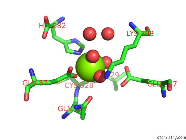
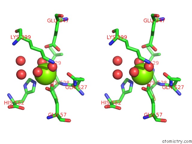
Magnesium binding site 1 out of 4 in 6g94
Go back to
Magnesium binding site 1 out
of 4 in the Structure of E. Coli Hydrogenase-1 C19G Variant in Complex with Cytochrome B
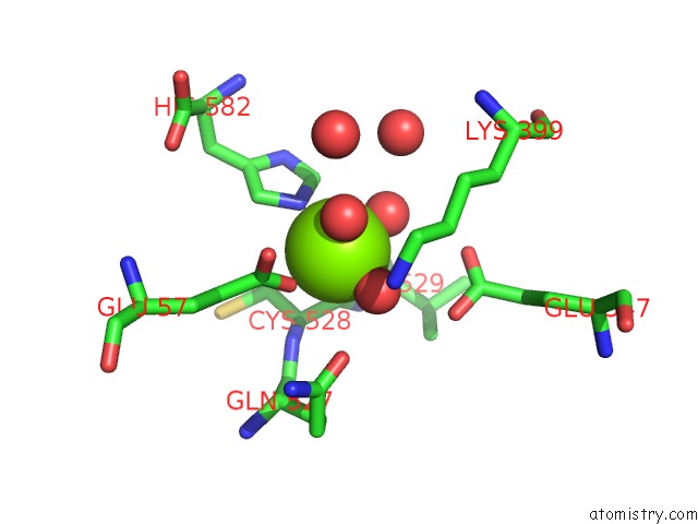
Mono view
Stereo pair view
Mono view
Stereo pair view
A full contact list of Magnesium with other atoms in the Mg binding
site number 1 of Structure of E. Coli Hydrogenase-1 C19G Variant in Complex with Cytochrome B within 5.0Å range:
|
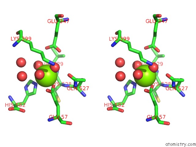
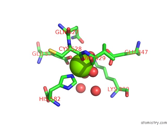
Magnesium binding site 2 out of 4 in 6g94
Go back to
Magnesium binding site 2 out
of 4 in the Structure of E. Coli Hydrogenase-1 C19G Variant in Complex with Cytochrome B
Mono view
Stereo pair view
Mono view
Stereo pair view
A full contact list of Magnesium with other atoms in the Mg binding
site number 2 of Structure of E. Coli Hydrogenase-1 C19G Variant in Complex with Cytochrome B within 5.0Å range:
|
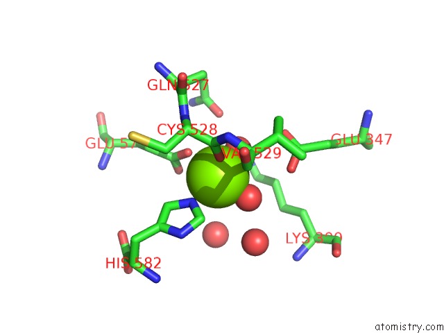
Magnesium binding site 3 out of 4 in 6g94
Go back to
Magnesium binding site 3 out
of 4 in the Structure of E. Coli Hydrogenase-1 C19G Variant in Complex with Cytochrome B
Mono view
Stereo pair view
Mono view
Stereo pair view
A full contact list of Magnesium with other atoms in the Mg binding
site number 3 of Structure of E. Coli Hydrogenase-1 C19G Variant in Complex with Cytochrome B within 5.0Å range:
|
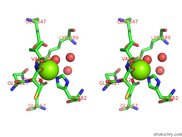
Magnesium binding site 4 out of 4 in 6g94
Go back to
Magnesium binding site 4 out
of 4 in the Structure of E. Coli Hydrogenase-1 C19G Variant in Complex with Cytochrome B
Mono view

Stereo pair view
Mono view
Stereo pair view
A full contact list of Magnesium with other atoms in the Mg binding
site number 4 of Structure of E. Coli Hydrogenase-1 C19G Variant in Complex with Cytochrome B within 5.0Å range:
|
Reference:
A.Volbeda,
J.M.Mouesca,
C.Darnault,
M.M.Roessler,
A.Parkin,
F.A.Armstrong,
J.C.Fontecilla-Camps.
X-Ray Structural, Functional and Computational Studies of the O2-Sensitive E. Coli Hydrogenase-1 C19G Variant Reveal An Unusual [4FE-4S] Cluster. Chem. Commun. (Camb.) V. 54 7175 2018.
ISSN: ESSN 1364-548X
PubMed: 29888350
DOI: 10.1039/C8CC02896F
Page generated: Tue Oct 1 01:03:09 2024
ISSN: ESSN 1364-548X
PubMed: 29888350
DOI: 10.1039/C8CC02896F
Last articles
Zn in 9MJ5Zn in 9HNW
Zn in 9G0L
Zn in 9FNE
Zn in 9DZN
Zn in 9E0I
Zn in 9D32
Zn in 9DAK
Zn in 8ZXC
Zn in 8ZUF