Magnesium in PDB 6gal: Structure of Fully Reduced Hydrogenase (Hyd-1) Variant E28Q Collected at pH 10
Enzymatic activity of Structure of Fully Reduced Hydrogenase (Hyd-1) Variant E28Q Collected at pH 10
All present enzymatic activity of Structure of Fully Reduced Hydrogenase (Hyd-1) Variant E28Q Collected at pH 10:
1.12.99.6;
Protein crystallography data
The structure of Structure of Fully Reduced Hydrogenase (Hyd-1) Variant E28Q Collected at pH 10, PDB code: 6gal
was solved by
S.B.Carr,
F.A.Armstrong,
R.M.Evans,
with X-Ray Crystallography technique. A brief refinement statistics is given in the table below:
Resolution Low / High (Å)
|
92.12 /
1.25
|
Space group
|
P 21 21 21
|
Cell size a, b, c (Å), α, β, γ (°)
|
94.906,
95.224,
183.910,
90.00,
90.00,
90.00
|
R / Rfree (%)
|
12 /
14.7
|
Other elements in 6gal:
The structure of Structure of Fully Reduced Hydrogenase (Hyd-1) Variant E28Q Collected at pH 10 also contains other interesting chemical elements:
Magnesium Binding Sites:
The binding sites of Magnesium atom in the Structure of Fully Reduced Hydrogenase (Hyd-1) Variant E28Q Collected at pH 10
(pdb code 6gal). This binding sites where shown within
5.0 Angstroms radius around Magnesium atom.
In total 2 binding sites of Magnesium where determined in the
Structure of Fully Reduced Hydrogenase (Hyd-1) Variant E28Q Collected at pH 10, PDB code: 6gal:
Jump to Magnesium binding site number:
1;
2;
Magnesium binding site 1 out
of 2 in 6gal
Go back to
Magnesium Binding Sites List in 6gal
Magnesium binding site 1 out
of 2 in the Structure of Fully Reduced Hydrogenase (Hyd-1) Variant E28Q Collected at pH 10
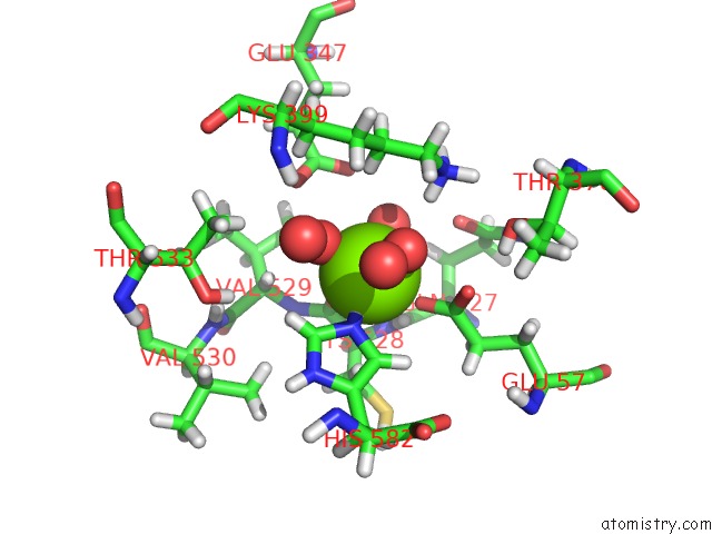
 Mono view
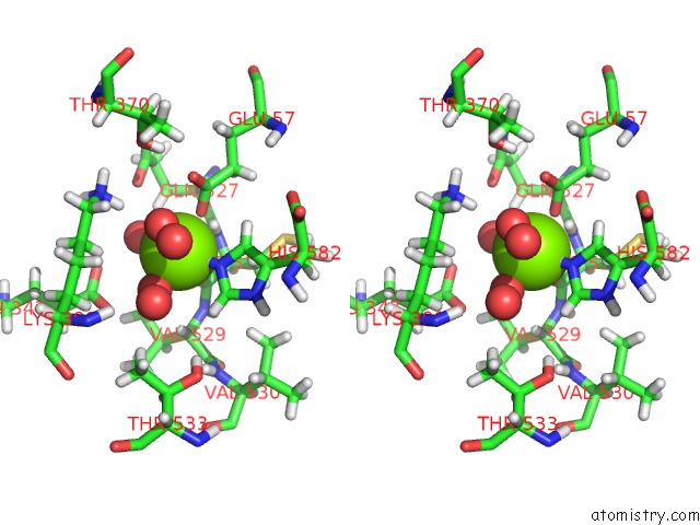
 Stereo pair view
|
A full contact list of Magnesium with other atoms in the Mg binding
site number 1 of Structure of Fully Reduced Hydrogenase (Hyd-1) Variant E28Q Collected at pH 10 within 5.0Å range:
probe
|
atom
|
residue
|
distance (Å)
|
B
|
Occ
|
L:Mg602
b:4.1
occ:1.00
|
HE2
|
L:HIS582
|
1.3
|
5.2
|
0.0
|
O
|
L:HOH725
|
2.1
|
6.4
|
1.0
|
O
|
L:HOH711
|
2.1
|
6.6
|
1.0
|
O
|
L:CYS528
|
2.1
|
5.9
|
1.0
|
O
|
L:HOH726
|
2.1
|
5.8
|
1.0
|
OE1
|
L:GLU57
|
2.2
|
6.1
|
1.0
|
NE2
|
L:HIS582
|
2.2
|
5.4
|
1.0
|
CD
|
L:GLU57
|
3.1
|
5.8
|
1.0
|
H
|
L:CYS528
|
3.1
|
5.0
|
1.0
|
CE1
|
L:HIS582
|
3.1
|
5.5
|
1.0
|
CD2
|
L:HIS582
|
3.2
|
6.2
|
1.0
|
HE1
|
L:HIS582
|
3.3
|
5.3
|
1.0
|
C
|
L:CYS528
|
3.3
|
5.1
|
1.0
|
HZ1
|
L:LYS399
|
3.3
|
6.6
|
1.0
|
HD2
|
L:HIS582
|
3.4
|
5.8
|
1.0
|
OE2
|
L:GLU57
|
3.4
|
6.7
|
1.0
|
HB2
|
L:CYS528
|
3.7
|
6.3
|
1.0
|
HD2
|
L:LYS399
|
3.7
|
6.9
|
1.0
|
N
|
L:CYS528
|
3.8
|
5.2
|
1.0
|
HA
|
L:VAL529
|
4.0
|
5.7
|
1.0
|
CA
|
L:CYS528
|
4.0
|
5.7
|
1.0
|
OE2
|
L:GLU347
|
4.1
|
7.0
|
1.0
|
OE1
|
L:GLU347
|
4.2
|
7.9
|
1.0
|
HG21
|
L:VAL529
|
4.2
|
7.0
|
1.0
|
OE1
|
L:GLN527
|
4.2
|
8.3
|
1.0
|
O
|
L:HOH1014
|
4.2
|
6.1
|
1.0
|
NZ
|
L:LYS399
|
4.3
|
6.5
|
1.0
|
ND1
|
L:HIS582
|
4.3
|
5.9
|
1.0
|
O
|
L:HOH828
|
4.3
|
6.5
|
1.0
|
CB
|
L:CYS528
|
4.3
|
6.3
|
1.0
|
CG
|
L:HIS582
|
4.3
|
5.7
|
1.0
|
N
|
L:VAL529
|
4.4
|
5.5
|
1.0
|
CG
|
L:GLU57
|
4.4
|
6.7
|
1.0
|
HE3
|
L:LYS399
|
4.5
|
6.4
|
1.0
|
CD
|
L:LYS399
|
4.5
|
7.1
|
1.0
|
CD
|
L:GLU347
|
4.6
|
7.2
|
1.0
|
HG2
|
L:GLU57
|
4.6
|
6.5
|
1.0
|
HG3
|
L:GLU57
|
4.6
|
7.0
|
1.0
|
HG1
|
L:THR370
|
4.6
|
7.0
|
0.0
|
CA
|
L:VAL529
|
4.7
|
5.8
|
1.0
|
HA
|
L:GLN527
|
4.7
|
5.7
|
1.0
|
CE
|
L:LYS399
|
4.7
|
6.4
|
1.0
|
HZ3
|
L:LYS399
|
4.7
|
6.6
|
1.0
|
HZ2
|
L:LYS399
|
4.8
|
6.5
|
1.0
|
HB3
|
L:GLN527
|
4.8
|
5.8
|
1.0
|
HG22
|
L:THR533
|
4.8
|
5.8
|
1.0
|
HG23
|
L:VAL530
|
4.9
|
7.1
|
1.0
|
HG3
|
L:LYS399
|
4.9
|
7.4
|
1.0
|
HB3
|
L:CYS528
|
4.9
|
6.3
|
1.0
|
HG1
|
L:THR533
|
4.9
|
6.1
|
0.0
|
C
|
L:GLN527
|
4.9
|
5.4
|
1.0
|
HA
|
L:CYS528
|
4.9
|
5.6
|
1.0
|
|
Magnesium binding site 2 out
of 2 in 6gal
Go back to
Magnesium Binding Sites List in 6gal
Magnesium binding site 2 out
of 2 in the Structure of Fully Reduced Hydrogenase (Hyd-1) Variant E28Q Collected at pH 10
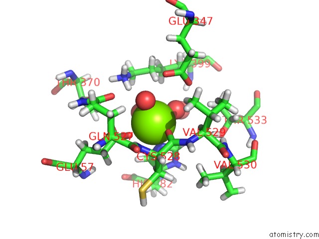
 Mono view
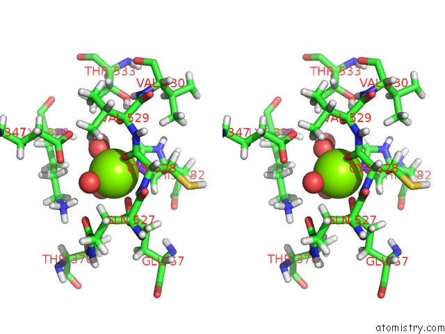
 Stereo pair view
|
A full contact list of Magnesium with other atoms in the Mg binding
site number 2 of Structure of Fully Reduced Hydrogenase (Hyd-1) Variant E28Q Collected at pH 10 within 5.0Å range:
probe
|
atom
|
residue
|
distance (Å)
|
B
|
Occ
|
M:Mg603
b:5.4
occ:1.00
|
HE2
|
M:HIS582
|
1.3
|
6.3
|
0.0
|
O
|
M:HOH719
|
2.1
|
7.5
|
1.0
|
O
|
M:HOH716
|
2.1
|
8.1
|
1.0
|
O
|
M:CYS528
|
2.1
|
7.7
|
1.0
|
O
|
M:HOH720
|
2.1
|
7.9
|
1.0
|
OE1
|
M:GLU57
|
2.2
|
6.8
|
1.0
|
NE2
|
M:HIS582
|
2.2
|
6.5
|
1.0
|
CE1
|
M:HIS582
|
3.1
|
6.3
|
1.0
|
CD
|
M:GLU57
|
3.1
|
7.2
|
1.0
|
H
|
M:CYS528
|
3.1
|
6.8
|
1.0
|
HE1
|
M:HIS582
|
3.2
|
6.3
|
1.0
|
CD2
|
M:HIS582
|
3.2
|
7.5
|
1.0
|
C
|
M:CYS528
|
3.3
|
6.6
|
1.0
|
HZ1
|
M:LYS399
|
3.3
|
8.2
|
1.0
|
OE2
|
M:GLU57
|
3.4
|
7.5
|
1.0
|
HD2
|
M:HIS582
|
3.4
|
7.0
|
1.0
|
HD2
|
M:LYS399
|
3.6
|
8.0
|
1.0
|
HB2
|
M:CYS528
|
3.7
|
7.4
|
1.0
|
N
|
M:CYS528
|
3.8
|
6.8
|
1.0
|
CA
|
M:CYS528
|
4.0
|
6.5
|
1.0
|
HA
|
M:VAL529
|
4.0
|
7.5
|
1.0
|
OE2
|
M:GLU347
|
4.1
|
9.0
|
1.0
|
OE1
|
M:GLU347
|
4.2
|
8.7
|
1.0
|
OE1
|
M:GLN527
|
4.2
|
9.5
|
1.0
|
HG21
|
M:VAL529
|
4.2
|
9.3
|
1.0
|
ND1
|
M:HIS582
|
4.3
|
6.5
|
1.0
|
O
|
M:HOH969
|
4.3
|
7.6
|
1.0
|
NZ
|
M:LYS399
|
4.3
|
8.1
|
1.0
|
O
|
M:HOH782
|
4.3
|
7.6
|
1.0
|
CB
|
M:CYS528
|
4.3
|
7.4
|
1.0
|
CG
|
M:HIS582
|
4.3
|
6.6
|
1.0
|
N
|
M:VAL529
|
4.4
|
6.9
|
1.0
|
CG
|
M:GLU57
|
4.4
|
7.0
|
1.0
|
HE3
|
M:LYS399
|
4.5
|
7.7
|
1.0
|
CD
|
M:LYS399
|
4.5
|
7.9
|
1.0
|
CD
|
M:GLU347
|
4.5
|
8.8
|
1.0
|
HG2
|
M:GLU57
|
4.6
|
7.2
|
1.0
|
HG3
|
M:GLU57
|
4.6
|
6.9
|
1.0
|
HG1
|
M:THR370
|
4.6
|
8.1
|
0.0
|
HA
|
M:GLN527
|
4.7
|
7.9
|
1.0
|
CA
|
M:VAL529
|
4.7
|
7.4
|
1.0
|
CE
|
M:LYS399
|
4.7
|
7.9
|
1.0
|
HZ3
|
M:LYS399
|
4.8
|
8.1
|
1.0
|
HZ2
|
M:LYS399
|
4.8
|
8.1
|
1.0
|
HB3
|
M:GLN527
|
4.8
|
8.3
|
1.0
|
HG22
|
M:THR533
|
4.8
|
6.5
|
1.0
|
HB3
|
M:CYS528
|
4.8
|
7.4
|
1.0
|
HG3
|
M:LYS399
|
4.9
|
8.4
|
1.0
|
HG23
|
M:VAL530
|
4.9
|
8.0
|
1.0
|
C
|
M:GLN527
|
4.9
|
7.0
|
1.0
|
HA
|
M:CYS528
|
4.9
|
6.7
|
1.0
|
HG1
|
M:THR533
|
4.9
|
6.6
|
0.0
|
|
Reference:
R.M.Evans,
P.A.Ash,
S.E.Beaton,
E.J.Brooke,
K.A.Vincent,
S.B.Carr,
F.A.Armstrong.
Mechanistic Exploitation of A Self-Repairing, Blocked Proton Transfer Pathway in An O2-Tolerant [Nife]-Hydrogenase. J. Am. Chem. Soc. V. 140 10208 2018.
ISSN: ESSN 1520-5126
PubMed: 30070475
DOI: 10.1021/JACS.8B04798
Page generated: Tue Oct 1 01:03:23 2024
|