Magnesium »
PDB 6g6z-6gfu »
6gfq »
Magnesium in PDB 6gfq: Cyanobacterial Gapdh with Nad and CP12 Bound
Protein crystallography data
The structure of Cyanobacterial Gapdh with Nad and CP12 Bound, PDB code: 6gfq
was solved by
C.R.Mcfarlane,
J.W.Murray,
with X-Ray Crystallography technique. A brief refinement statistics is given in the table below:
| Resolution Low / High (Å) | 41.77 / 1.40 |
| Space group | P 43 21 2 |
| Cell size a, b, c (Å), α, β, γ (°) | 141.217, 141.217, 76.238, 90.00, 90.00, 90.00 |
| R / Rfree (%) | 14.6 / 17.4 |
Magnesium Binding Sites:
The binding sites of Magnesium atom in the Cyanobacterial Gapdh with Nad and CP12 Bound
(pdb code 6gfq). This binding sites where shown within
5.0 Angstroms radius around Magnesium atom.
In total 2 binding sites of Magnesium where determined in the Cyanobacterial Gapdh with Nad and CP12 Bound, PDB code: 6gfq:
Jump to Magnesium binding site number: 1; 2;
In total 2 binding sites of Magnesium where determined in the Cyanobacterial Gapdh with Nad and CP12 Bound, PDB code: 6gfq:
Jump to Magnesium binding site number: 1; 2;
Magnesium binding site 1 out of 2 in 6gfq
Go back to
Magnesium binding site 1 out
of 2 in the Cyanobacterial Gapdh with Nad and CP12 Bound
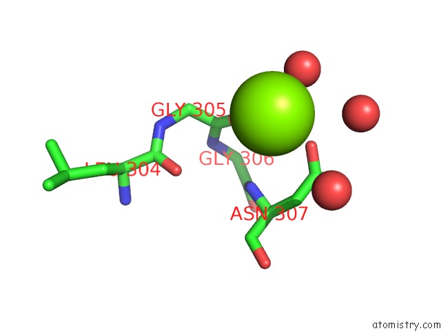
Mono view
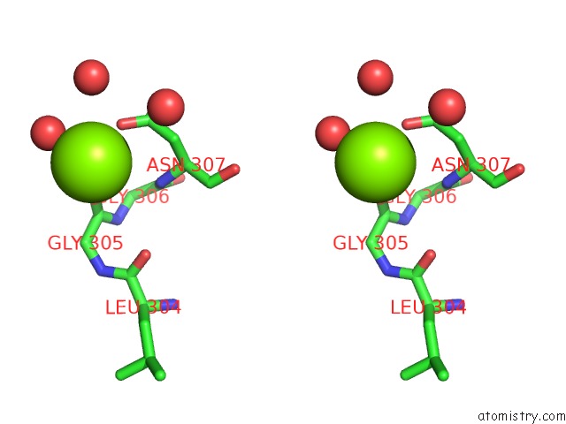
Stereo pair view
Mono view
Stereo pair view
A full contact list of Magnesium with other atoms in the Mg binding
site number 1 of Cyanobacterial Gapdh with Nad and CP12 Bound within 5.0Å range:
|
Magnesium binding site 2 out of 2 in 6gfq
Go back to
Magnesium binding site 2 out
of 2 in the Cyanobacterial Gapdh with Nad and CP12 Bound
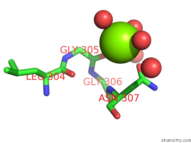
Mono view
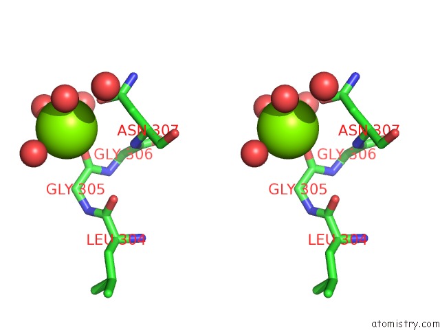
Stereo pair view
Mono view
Stereo pair view
A full contact list of Magnesium with other atoms in the Mg binding
site number 2 of Cyanobacterial Gapdh with Nad and CP12 Bound within 5.0Å range:
|
Reference:
C.R.Mcfarlane,
N.R.Shah,
B.V.Kabasakal,
B.Echeverria,
C.A.R.Cotton,
D.Bubeck,
J.W.Murray.
Structural Basis of Light-Induced Redox Regulation in the Calvin-Benson Cycle in Cyanobacteria. Proc.Natl.Acad.Sci.Usa V. 116 20984 2019.
ISSN: ESSN 1091-6490
PubMed: 31570616
DOI: 10.1073/PNAS.1906722116
Page generated: Tue Oct 1 01:09:15 2024
ISSN: ESSN 1091-6490
PubMed: 31570616
DOI: 10.1073/PNAS.1906722116
Last articles
Zn in 9J0NZn in 9J0O
Zn in 9J0P
Zn in 9FJX
Zn in 9EKB
Zn in 9C0F
Zn in 9CAH
Zn in 9CH0
Zn in 9CH3
Zn in 9CH1