Magnesium »
PDB 6gg6-6gof »
6ghb »
Magnesium in PDB 6ghb: Crystal Structure of Spx in Complex with Yjbh (Oxidized)
Protein crystallography data
The structure of Crystal Structure of Spx in Complex with Yjbh (Oxidized), PDB code: 6ghb
was solved by
W.Awad,
D.T.Logan,
C.Von Wachenfeldt,
with X-Ray Crystallography technique. A brief refinement statistics is given in the table below:
| Resolution Low / High (Å) | 48.43 / 3.10 |
| Space group | P 21 21 21 |
| Cell size a, b, c (Å), α, β, γ (°) | 85.596, 88.704, 117.467, 90.00, 90.00, 90.00 |
| R / Rfree (%) | 22.2 / 26.6 |
Magnesium Binding Sites:
The binding sites of Magnesium atom in the Crystal Structure of Spx in Complex with Yjbh (Oxidized)
(pdb code 6ghb). This binding sites where shown within
5.0 Angstroms radius around Magnesium atom.
In total 2 binding sites of Magnesium where determined in the Crystal Structure of Spx in Complex with Yjbh (Oxidized), PDB code: 6ghb:
Jump to Magnesium binding site number: 1; 2;
In total 2 binding sites of Magnesium where determined in the Crystal Structure of Spx in Complex with Yjbh (Oxidized), PDB code: 6ghb:
Jump to Magnesium binding site number: 1; 2;
Magnesium binding site 1 out of 2 in 6ghb
Go back to
Magnesium binding site 1 out
of 2 in the Crystal Structure of Spx in Complex with Yjbh (Oxidized)
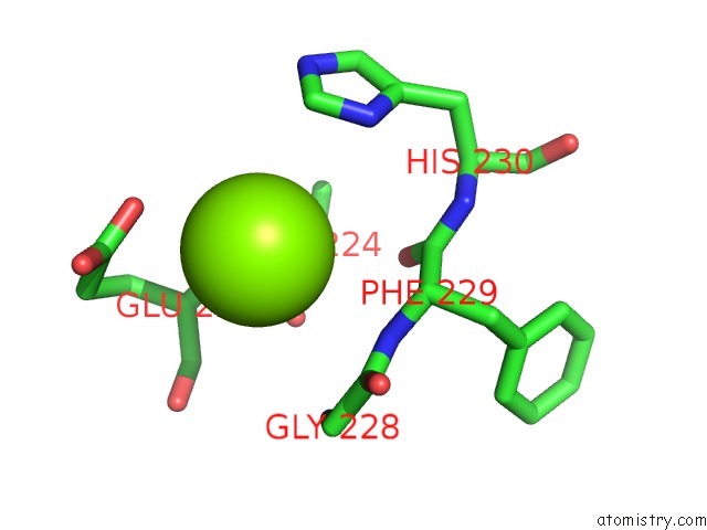
Mono view
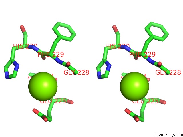
Stereo pair view
Mono view
Stereo pair view
A full contact list of Magnesium with other atoms in the Mg binding
site number 1 of Crystal Structure of Spx in Complex with Yjbh (Oxidized) within 5.0Å range:
|
Magnesium binding site 2 out of 2 in 6ghb
Go back to
Magnesium binding site 2 out
of 2 in the Crystal Structure of Spx in Complex with Yjbh (Oxidized)
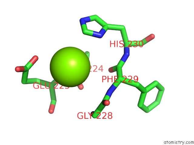
Mono view
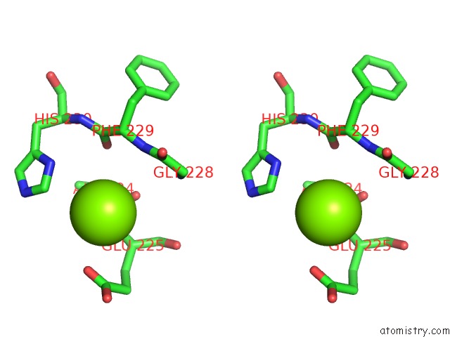
Stereo pair view
Mono view
Stereo pair view
A full contact list of Magnesium with other atoms in the Mg binding
site number 2 of Crystal Structure of Spx in Complex with Yjbh (Oxidized) within 5.0Å range:
|
Reference:
W.Awad,
Y.Al-Eryani,
S.Ekstrom,
D.T.Logan,
C.Von Wachenfeldt.
Structural Basis For Yjbh Adaptor-Mediated Recognition of Transcription Factor Spx. Structure V. 27 923 2019.
ISSN: ISSN 0969-2126
PubMed: 30982633
DOI: 10.1016/J.STR.2019.03.009
Page generated: Tue Oct 1 01:10:24 2024
ISSN: ISSN 0969-2126
PubMed: 30982633
DOI: 10.1016/J.STR.2019.03.009
Last articles
Zn in 9JYWZn in 9IR4
Zn in 9IR3
Zn in 9GMX
Zn in 9GMW
Zn in 9JEJ
Zn in 9ERF
Zn in 9ERE
Zn in 9EGV
Zn in 9EGW