Magnesium »
PDB 6gom-6gtr »
6gs2 »
Magnesium in PDB 6gs2: Crystal Structure of the Gatd/Murt Enzyme Complex From Staphylococcus Aureus
Protein crystallography data
The structure of Crystal Structure of the Gatd/Murt Enzyme Complex From Staphylococcus Aureus, PDB code: 6gs2
was solved by
L.M.Muckenfuss,
E.R.Noeldeke,
V.Niemann,
G.Zocher,
T.Stehle,
with X-Ray Crystallography technique. A brief refinement statistics is given in the table below:
| Resolution Low / High (Å) | 49.06 / 2.04 |
| Space group | P 21 21 21 |
| Cell size a, b, c (Å), α, β, γ (°) | 107.100, 110.370, 116.360, 90.00, 90.00, 90.00 |
| R / Rfree (%) | 17.5 / 21.6 |
Other elements in 6gs2:
The structure of Crystal Structure of the Gatd/Murt Enzyme Complex From Staphylococcus Aureus also contains other interesting chemical elements:
| Zinc | (Zn) | 2 atoms |
Magnesium Binding Sites:
The binding sites of Magnesium atom in the Crystal Structure of the Gatd/Murt Enzyme Complex From Staphylococcus Aureus
(pdb code 6gs2). This binding sites where shown within
5.0 Angstroms radius around Magnesium atom.
In total only one binding site of Magnesium was determined in the Crystal Structure of the Gatd/Murt Enzyme Complex From Staphylococcus Aureus, PDB code: 6gs2:
In total only one binding site of Magnesium was determined in the Crystal Structure of the Gatd/Murt Enzyme Complex From Staphylococcus Aureus, PDB code: 6gs2:
Magnesium binding site 1 out of 1 in 6gs2
Go back to
Magnesium binding site 1 out
of 1 in the Crystal Structure of the Gatd/Murt Enzyme Complex From Staphylococcus Aureus
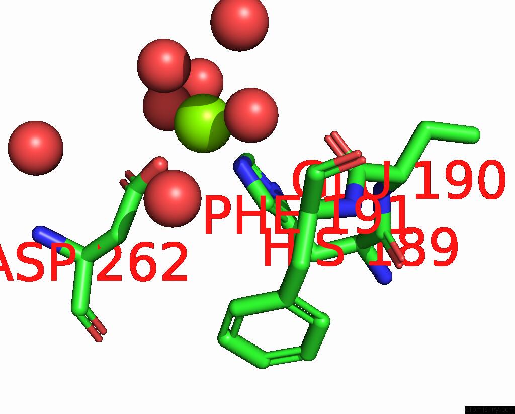
Mono view
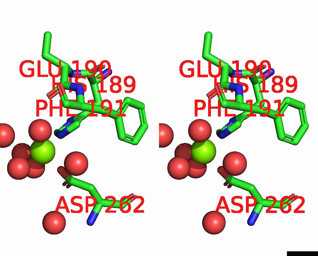
Stereo pair view
Mono view
Stereo pair view
A full contact list of Magnesium with other atoms in the Mg binding
site number 1 of Crystal Structure of the Gatd/Murt Enzyme Complex From Staphylococcus Aureus within 5.0Å range:
|
Reference:
E.R.Noldeke,
L.M.Muckenfuss,
V.Niemann,
A.Muller,
E.Stork,
G.Zocher,
T.Schneider,
T.Stehle.
Structural Basis of Cell Wall Peptidoglycan Amidation By the Gatd/Murt Complex of Staphylococcus Aureus. Sci Rep V. 8 12953 2018.
ISSN: ESSN 2045-2322
PubMed: 30154570
DOI: 10.1038/S41598-018-31098-X
Page generated: Tue Oct 1 01:24:07 2024
ISSN: ESSN 2045-2322
PubMed: 30154570
DOI: 10.1038/S41598-018-31098-X
Last articles
Zn in 9MJ5Zn in 9HNW
Zn in 9G0L
Zn in 9FNE
Zn in 9DZN
Zn in 9E0I
Zn in 9D32
Zn in 9DAK
Zn in 8ZXC
Zn in 8ZUF