Magnesium »
PDB 6guj-6h57 »
6gw9 »
Magnesium in PDB 6gw9: Concanavalin A Structure Determined with Data From the Euxfel, the First Mhz Free Electron Laser
Protein crystallography data
The structure of Concanavalin A Structure Determined with Data From the Euxfel, the First Mhz Free Electron Laser, PDB code: 6gw9
was solved by
M.L.Gruenbein,
A.Gorel,
M.Stricker,
R.Bean,
J.Bielecki,
K.Doerner,
E.Hartmann,
M.Hilpert,
M.Kloos,
R.Letrun,
J.Sztuk-Dambietz,
A.Mancuso,
M.Meserschmidt,
G.Nass-Kovacs,
M.Ramilli,
C.M.Roome,
T.Sato,
R.B.Doak,
R.L.Shoeman,
L.Foucar,
J.P.Colletier,
T.R.M.Barends,
C.Stan,
I.Schlichting,
with X-Ray Crystallography technique. A brief refinement statistics is given in the table below:
| Resolution Low / High (Å) | 45.00 / 2.10 |
| Space group | I 2 2 2 |
| Cell size a, b, c (Å), α, β, γ (°) | 63.800, 88.100, 90.200, 90.00, 90.00, 90.00 |
| R / Rfree (%) | 18.6 / 23.8 |
Other elements in 6gw9:
The structure of Concanavalin A Structure Determined with Data From the Euxfel, the First Mhz Free Electron Laser also contains other interesting chemical elements:
| Calcium | (Ca) | 1 atom |
Magnesium Binding Sites:
The binding sites of Magnesium atom in the Concanavalin A Structure Determined with Data From the Euxfel, the First Mhz Free Electron Laser
(pdb code 6gw9). This binding sites where shown within
5.0 Angstroms radius around Magnesium atom.
In total only one binding site of Magnesium was determined in the Concanavalin A Structure Determined with Data From the Euxfel, the First Mhz Free Electron Laser, PDB code: 6gw9:
In total only one binding site of Magnesium was determined in the Concanavalin A Structure Determined with Data From the Euxfel, the First Mhz Free Electron Laser, PDB code: 6gw9:
Magnesium binding site 1 out of 1 in 6gw9
Go back to
Magnesium binding site 1 out
of 1 in the Concanavalin A Structure Determined with Data From the Euxfel, the First Mhz Free Electron Laser
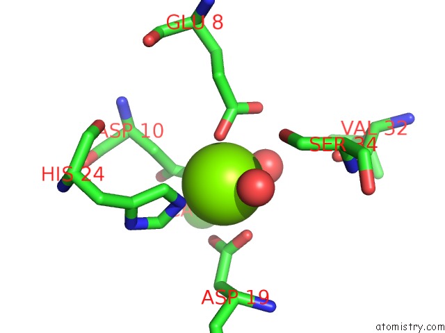
Mono view
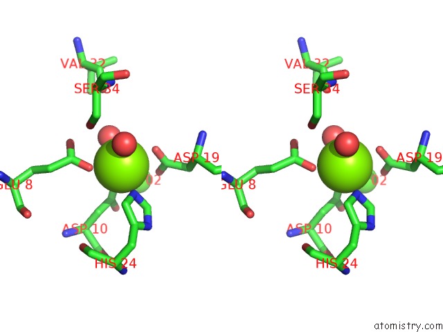
Stereo pair view
Mono view
Stereo pair view
A full contact list of Magnesium with other atoms in the Mg binding
site number 1 of Concanavalin A Structure Determined with Data From the Euxfel, the First Mhz Free Electron Laser within 5.0Å range:
|
Reference:
M.L.Grunbein,
J.Bielecki,
A.Gorel,
M.Stricker,
R.Bean,
M.Cammarata,
K.Dorner,
L.Frohlich,
E.Hartmann,
S.Hauf,
M.Hilpert,
Y.Kim,
M.Kloos,
R.Letrun,
M.Messerschmidt,
G.Mills,
G.Nass Kovacs,
M.Ramilli,
C.M.Roome,
T.Sato,
M.Scholz,
M.Sliwa,
J.Sztuk-Dambietz,
M.Weik,
B.Weinhausen,
N.Al-Qudami,
D.Boukhelef,
S.Brockhauser,
W.Ehsan,
M.Emons,
S.Esenov,
H.Fangohr,
A.Kaukher,
T.Kluyver,
M.Lederer,
L.Maia,
M.Manetti,
T.Michelat,
A.Munnich,
F.Pallas,
G.Palmer,
G.Previtali,
N.Raab,
A.Silenzi,
J.Szuba,
S.Venkatesan,
K.Wrona,
J.Zhu,
R.B.Doak,
R.L.Shoeman,
L.Foucar,
J.P.Colletier,
A.P.Mancuso,
T.R.M.Barends,
C.A.Stan,
I.Schlichting.
Megahertz Data Collection From Protein Microcrystals at An X-Ray Free-Electron Laser. Nat Commun V. 9 3487 2018.
ISSN: ESSN 2041-1723
PubMed: 30154468
DOI: 10.1038/S41467-018-05953-4
Page generated: Tue Oct 1 01:26:03 2024
ISSN: ESSN 2041-1723
PubMed: 30154468
DOI: 10.1038/S41467-018-05953-4
Last articles
Zn in 9MJ5Zn in 9HNW
Zn in 9G0L
Zn in 9FNE
Zn in 9DZN
Zn in 9E0I
Zn in 9D32
Zn in 9DAK
Zn in 8ZXC
Zn in 8ZUF