Magnesium »
PDB 6h57-6hcn »
6h99 »
Magnesium in PDB 6h99: Crystal Structure of Anaerobic Ergothioneine Biosynthesis Enzyme From Chlorobium Limicola in Persulfide Form.
Protein crystallography data
The structure of Crystal Structure of Anaerobic Ergothioneine Biosynthesis Enzyme From Chlorobium Limicola in Persulfide Form., PDB code: 6h99
was solved by
F.Leisinger,
R.Burn,
M.Meury,
P.Lukat,
F.P.Seebeck,
with X-Ray Crystallography technique. A brief refinement statistics is given in the table below:
| Resolution Low / High (Å) | 43.54 / 1.60 |
| Space group | C 1 2 1 |
| Cell size a, b, c (Å), α, β, γ (°) | 129.760, 43.183, 88.740, 90.00, 108.46, 90.00 |
| R / Rfree (%) | 16.3 / 19.5 |
Other elements in 6h99:
The structure of Crystal Structure of Anaerobic Ergothioneine Biosynthesis Enzyme From Chlorobium Limicola in Persulfide Form. also contains other interesting chemical elements:
| Chlorine | (Cl) | 4 atoms |
Magnesium Binding Sites:
The binding sites of Magnesium atom in the Crystal Structure of Anaerobic Ergothioneine Biosynthesis Enzyme From Chlorobium Limicola in Persulfide Form.
(pdb code 6h99). This binding sites where shown within
5.0 Angstroms radius around Magnesium atom.
In total 4 binding sites of Magnesium where determined in the Crystal Structure of Anaerobic Ergothioneine Biosynthesis Enzyme From Chlorobium Limicola in Persulfide Form., PDB code: 6h99:
Jump to Magnesium binding site number: 1; 2; 3; 4;
In total 4 binding sites of Magnesium where determined in the Crystal Structure of Anaerobic Ergothioneine Biosynthesis Enzyme From Chlorobium Limicola in Persulfide Form., PDB code: 6h99:
Jump to Magnesium binding site number: 1; 2; 3; 4;
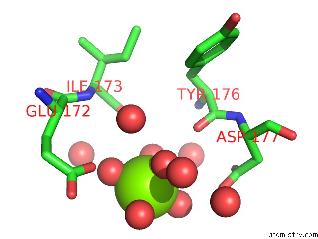
Magnesium binding site 1 out of 4 in 6h99
Go back to
Magnesium binding site 1 out
of 4 in the Crystal Structure of Anaerobic Ergothioneine Biosynthesis Enzyme From Chlorobium Limicola in Persulfide Form.

Mono view

Stereo pair view
Mono view
Stereo pair view
A full contact list of Magnesium with other atoms in the Mg binding
site number 1 of Crystal Structure of Anaerobic Ergothioneine Biosynthesis Enzyme From Chlorobium Limicola in Persulfide Form. within 5.0Å range:
|
Magnesium binding site 2 out of 4 in 6h99
Go back to
Magnesium binding site 2 out
of 4 in the Crystal Structure of Anaerobic Ergothioneine Biosynthesis Enzyme From Chlorobium Limicola in Persulfide Form.

Mono view

Stereo pair view
Mono view
Stereo pair view
A full contact list of Magnesium with other atoms in the Mg binding
site number 2 of Crystal Structure of Anaerobic Ergothioneine Biosynthesis Enzyme From Chlorobium Limicola in Persulfide Form. within 5.0Å range:
|
Magnesium binding site 3 out of 4 in 6h99
Go back to
Magnesium binding site 3 out
of 4 in the Crystal Structure of Anaerobic Ergothioneine Biosynthesis Enzyme From Chlorobium Limicola in Persulfide Form.
Mono view

Stereo pair view
Mono view
Stereo pair view
A full contact list of Magnesium with other atoms in the Mg binding
site number 3 of Crystal Structure of Anaerobic Ergothioneine Biosynthesis Enzyme From Chlorobium Limicola in Persulfide Form. within 5.0Å range:
|
Magnesium binding site 4 out of 4 in 6h99
Go back to
Magnesium binding site 4 out
of 4 in the Crystal Structure of Anaerobic Ergothioneine Biosynthesis Enzyme From Chlorobium Limicola in Persulfide Form.

Mono view

Stereo pair view
Mono view
Stereo pair view
A full contact list of Magnesium with other atoms in the Mg binding
site number 4 of Crystal Structure of Anaerobic Ergothioneine Biosynthesis Enzyme From Chlorobium Limicola in Persulfide Form. within 5.0Å range:
|
Reference:
F.Leisinger,
R.Burn,
M.Meury,
P.Lukat,
F.P.Seebeck.
Structural and Mechanistic Basis For Anaerobic Ergothioneine Biosynthesis. J.Am.Chem.Soc. V. 141 6906 2019.
ISSN: ESSN 1520-5126
PubMed: 30943021
DOI: 10.1021/JACS.8B12596
Page generated: Tue Oct 1 01:38:02 2024
ISSN: ESSN 1520-5126
PubMed: 30943021
DOI: 10.1021/JACS.8B12596
Last articles
Zn in 9JYWZn in 9IR4
Zn in 9IR3
Zn in 9GMX
Zn in 9GMW
Zn in 9JEJ
Zn in 9ERF
Zn in 9ERE
Zn in 9EGV
Zn in 9EGW