Magnesium »
PDB 6hnq-6hvu »
6hq4 »
Magnesium in PDB 6hq4: Structure of Eal Enzyme BD1971 - Camp Bound Form
Protein crystallography data
The structure of Structure of Eal Enzyme BD1971 - Camp Bound Form, PDB code: 6hq4
was solved by
A.L.Lovering,
I.T.Cadby,
with X-Ray Crystallography technique. A brief refinement statistics is given in the table below:
| Resolution Low / High (Å) | 65.66 / 2.63 |
| Space group | P 31 |
| Cell size a, b, c (Å), α, β, γ (°) | 82.380, 82.380, 131.320, 90.00, 90.00, 120.00 |
| R / Rfree (%) | 24.1 / 28.6 |
Magnesium Binding Sites:
The binding sites of Magnesium atom in the Structure of Eal Enzyme BD1971 - Camp Bound Form
(pdb code 6hq4). This binding sites where shown within
5.0 Angstroms radius around Magnesium atom.
In total 2 binding sites of Magnesium where determined in the Structure of Eal Enzyme BD1971 - Camp Bound Form, PDB code: 6hq4:
Jump to Magnesium binding site number: 1; 2;
In total 2 binding sites of Magnesium where determined in the Structure of Eal Enzyme BD1971 - Camp Bound Form, PDB code: 6hq4:
Jump to Magnesium binding site number: 1; 2;
Magnesium binding site 1 out of 2 in 6hq4
Go back to
Magnesium binding site 1 out
of 2 in the Structure of Eal Enzyme BD1971 - Camp Bound Form
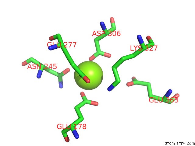
Mono view
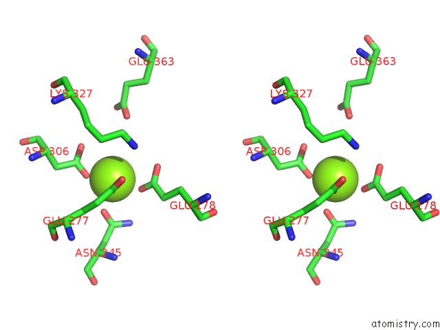
Stereo pair view
Mono view
Stereo pair view
A full contact list of Magnesium with other atoms in the Mg binding
site number 1 of Structure of Eal Enzyme BD1971 - Camp Bound Form within 5.0Å range:
|
Magnesium binding site 2 out of 2 in 6hq4
Go back to
Magnesium binding site 2 out
of 2 in the Structure of Eal Enzyme BD1971 - Camp Bound Form
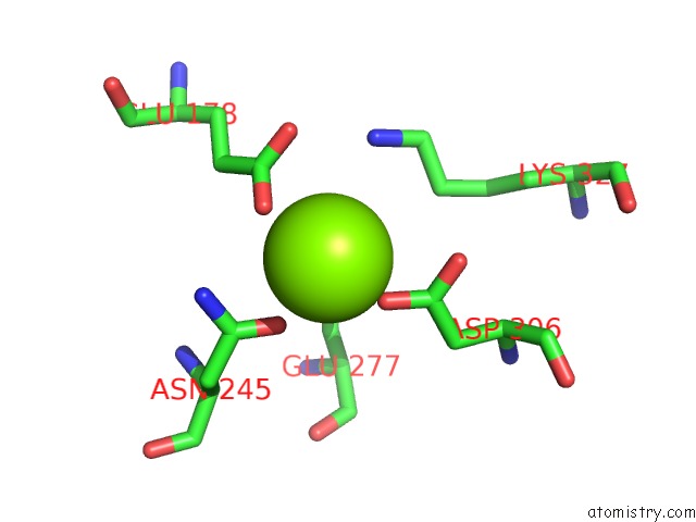
Mono view
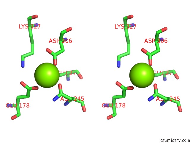
Stereo pair view
Mono view
Stereo pair view
A full contact list of Magnesium with other atoms in the Mg binding
site number 2 of Structure of Eal Enzyme BD1971 - Camp Bound Form within 5.0Å range:
|
Reference:
I.T.Cadby,
S.M.Basford,
R.Nottingham,
R.Meek,
R.Lowry,
C.Lambert,
M.Tridgett,
R.Till,
R.Ahmad,
R.Fung,
L.Hobley,
W.S.Hughes,
P.J.Moynihan,
R.E.Sockett,
A.L.Lovering.
Nucleotide Signaling Pathway Convergence in A Camp-Sensing Bacterial C-Di-Gmp Phosphodiesterase. Embo J. V. 38 00772 2019.
ISSN: ESSN 1460-2075
PubMed: 31355487
DOI: 10.15252/EMBJ.2018100772
Page generated: Tue Oct 1 02:11:42 2024
ISSN: ESSN 1460-2075
PubMed: 31355487
DOI: 10.15252/EMBJ.2018100772
Last articles
Zn in 9J0NZn in 9J0O
Zn in 9J0P
Zn in 9FJX
Zn in 9EKB
Zn in 9C0F
Zn in 9CAH
Zn in 9CH0
Zn in 9CH3
Zn in 9CH1