Magnesium »
PDB 6hns-6hvv »
6hrc »
Magnesium in PDB 6hrc: Outward-Facing Pglk with Atpgammas Bound
Protein crystallography data
The structure of Outward-Facing Pglk with Atpgammas Bound, PDB code: 6hrc
was solved by
C.Perez,
K.P.Locher,
with X-Ray Crystallography technique. A brief refinement statistics is given in the table below:
| Resolution Low / High (Å) | 20.00 / 3.30 |
| Space group | P 21 21 21 |
| Cell size a, b, c (Å), α, β, γ (°) | 125.680, 145.980, 206.760, 90.00, 90.00, 90.00 |
| R / Rfree (%) | 23.2 / 28.4 |
Magnesium Binding Sites:
The binding sites of Magnesium atom in the Outward-Facing Pglk with Atpgammas Bound
(pdb code 6hrc). This binding sites where shown within
5.0 Angstroms radius around Magnesium atom.
In total 2 binding sites of Magnesium where determined in the Outward-Facing Pglk with Atpgammas Bound, PDB code: 6hrc:
Jump to Magnesium binding site number: 1; 2;
In total 2 binding sites of Magnesium where determined in the Outward-Facing Pglk with Atpgammas Bound, PDB code: 6hrc:
Jump to Magnesium binding site number: 1; 2;
Magnesium binding site 1 out of 2 in 6hrc
Go back to
Magnesium binding site 1 out
of 2 in the Outward-Facing Pglk with Atpgammas Bound
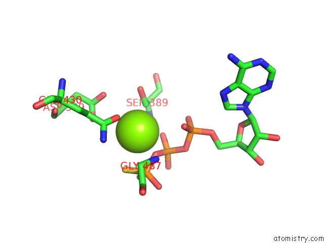
Mono view
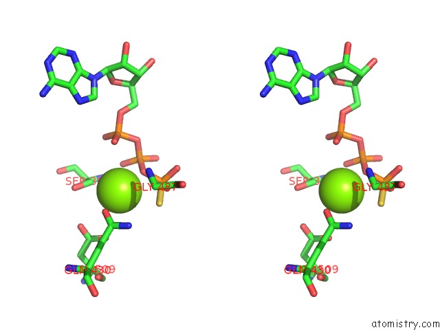
Stereo pair view
Mono view
Stereo pair view
A full contact list of Magnesium with other atoms in the Mg binding
site number 1 of Outward-Facing Pglk with Atpgammas Bound within 5.0Å range:
|
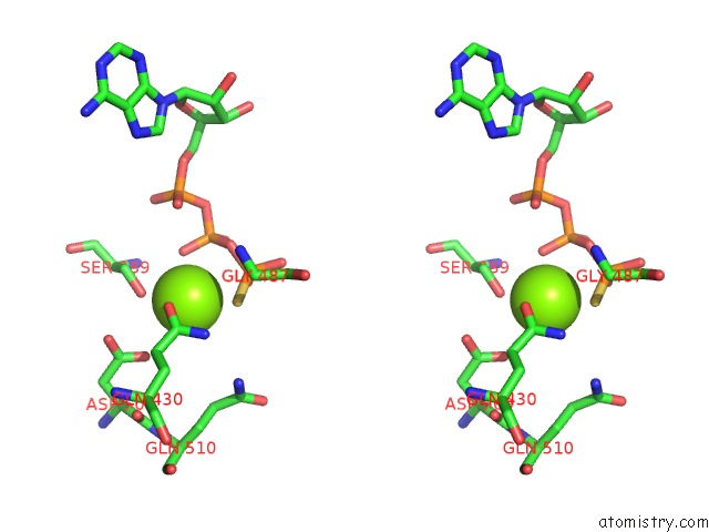
Magnesium binding site 2 out of 2 in 6hrc
Go back to
Magnesium binding site 2 out
of 2 in the Outward-Facing Pglk with Atpgammas Bound
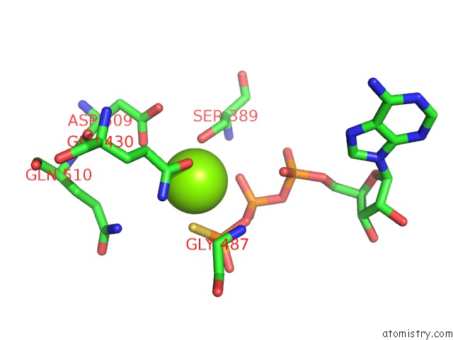
Mono view
Stereo pair view
Mono view
Stereo pair view
A full contact list of Magnesium with other atoms in the Mg binding
site number 2 of Outward-Facing Pglk with Atpgammas Bound within 5.0Å range:
|
Reference:
C.Perez,
A.R.Mehdipour,
G.Hummer,
K.P.Locher.
Structure of Outward-Facing Pglk and Molecular Dynamics of Lipid-Linked Oligosaccharide Recognition and Translocation. Structure V. 27 669 2019.
ISSN: ISSN 0969-2126
PubMed: 30799077
DOI: 10.1016/J.STR.2019.01.013
Page generated: Tue Oct 1 02:12:23 2024
ISSN: ISSN 0969-2126
PubMed: 30799077
DOI: 10.1016/J.STR.2019.01.013
Last articles
Cl in 6ALECl in 6AKI
Cl in 6AKW
Cl in 6AI5
Cl in 6AJI
Cl in 6AKV
Cl in 6AK8
Cl in 6AHS
Cl in 6AHL
Cl in 6AHY