Magnesium »
PDB 6i0v-6i9t »
6i3d »
Magnesium in PDB 6i3d: Crystal Structure of Human Soluble Catechol O-Methyltransferase in Complex with 3,5-Dinitrocatechol and Sinefungin
Enzymatic activity of Crystal Structure of Human Soluble Catechol O-Methyltransferase in Complex with 3,5-Dinitrocatechol and Sinefungin
All present enzymatic activity of Crystal Structure of Human Soluble Catechol O-Methyltransferase in Complex with 3,5-Dinitrocatechol and Sinefungin:
2.1.1.6;
2.1.1.6;
Protein crystallography data
The structure of Crystal Structure of Human Soluble Catechol O-Methyltransferase in Complex with 3,5-Dinitrocatechol and Sinefungin, PDB code: 6i3d
was solved by
C.W.Levy,
with X-Ray Crystallography technique. A brief refinement statistics is given in the table below:
| Resolution Low / High (Å) | 37.02 / 1.45 |
| Space group | P 1 21 1 |
| Cell size a, b, c (Å), α, β, γ (°) | 42.961, 75.797, 64.345, 90.00, 94.62, 90.00 |
| R / Rfree (%) | 11.9 / 14.6 |
Magnesium Binding Sites:
The binding sites of Magnesium atom in the Crystal Structure of Human Soluble Catechol O-Methyltransferase in Complex with 3,5-Dinitrocatechol and Sinefungin
(pdb code 6i3d). This binding sites where shown within
5.0 Angstroms radius around Magnesium atom.
In total 2 binding sites of Magnesium where determined in the Crystal Structure of Human Soluble Catechol O-Methyltransferase in Complex with 3,5-Dinitrocatechol and Sinefungin, PDB code: 6i3d:
Jump to Magnesium binding site number: 1; 2;
In total 2 binding sites of Magnesium where determined in the Crystal Structure of Human Soluble Catechol O-Methyltransferase in Complex with 3,5-Dinitrocatechol and Sinefungin, PDB code: 6i3d:
Jump to Magnesium binding site number: 1; 2;
Magnesium binding site 1 out of 2 in 6i3d
Go back to
Magnesium binding site 1 out
of 2 in the Crystal Structure of Human Soluble Catechol O-Methyltransferase in Complex with 3,5-Dinitrocatechol and Sinefungin
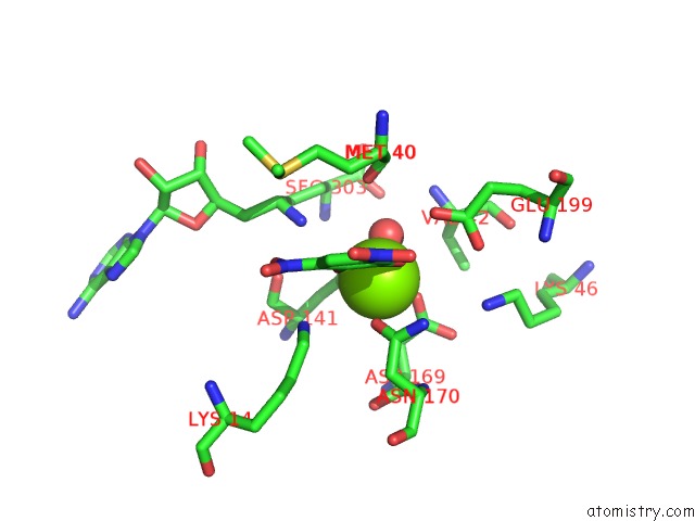
Mono view
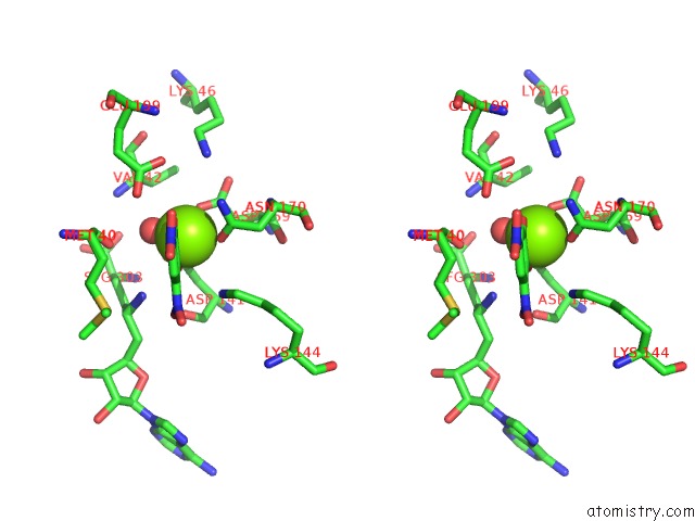
Stereo pair view
Mono view
Stereo pair view
A full contact list of Magnesium with other atoms in the Mg binding
site number 1 of Crystal Structure of Human Soluble Catechol O-Methyltransferase in Complex with 3,5-Dinitrocatechol and Sinefungin within 5.0Å range:
|
Magnesium binding site 2 out of 2 in 6i3d
Go back to
Magnesium binding site 2 out
of 2 in the Crystal Structure of Human Soluble Catechol O-Methyltransferase in Complex with 3,5-Dinitrocatechol and Sinefungin
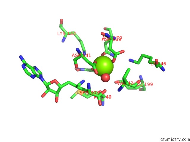
Mono view
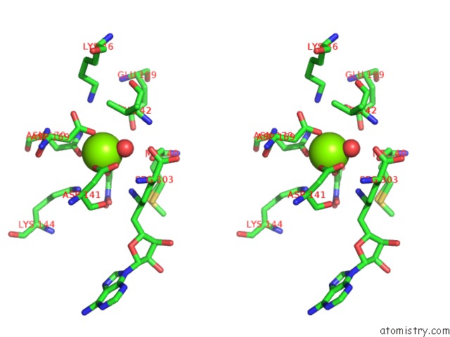
Stereo pair view
Mono view
Stereo pair view
A full contact list of Magnesium with other atoms in the Mg binding
site number 2 of Crystal Structure of Human Soluble Catechol O-Methyltransferase in Complex with 3,5-Dinitrocatechol and Sinefungin within 5.0Å range:
|
Reference:
S.Czarnota,
L.O.Johannissen,
N.J.Baxter,
F.Rummel,
A.L.Wilson,
M.J.Cliff,
C.W.Levy,
N.S.Scrutton,
J.P.Waltho,
S.Hay.
Equatorial Active Site Compaction and Electrostatic Reorganization in Catechol-O-Methyltransferase. Acs Catalysis V. 9 4394 2019.
ISSN: ESSN 2155-5435
PubMed: 31080692
DOI: 10.1021/ACSCATAL.9B00174
Page generated: Tue Oct 1 02:58:02 2024
ISSN: ESSN 2155-5435
PubMed: 31080692
DOI: 10.1021/ACSCATAL.9B00174
Last articles
Zn in 9J0NZn in 9J0O
Zn in 9J0P
Zn in 9FJX
Zn in 9EKB
Zn in 9C0F
Zn in 9CAH
Zn in 9CH0
Zn in 9CH3
Zn in 9CH1