Magnesium »
PDB 6i0v-6i9t »
6i5b »
Magnesium in PDB 6i5b: Crystal Structure of Outer Cell Wall Cytochrome Ocwa
Protein crystallography data
The structure of Crystal Structure of Outer Cell Wall Cytochrome Ocwa, PDB code: 6i5b
was solved by
B.Hermann,
O.Einsle,
with X-Ray Crystallography technique. A brief refinement statistics is given in the table below:
| Resolution Low / High (Å) | 80.94 / 2.20 |
| Space group | P 1 |
| Cell size a, b, c (Å), α, β, γ (°) | 54.855, 62.969, 84.210, 101.54, 99.13, 98.41 |
| R / Rfree (%) | 20.3 / 23.3 |
Other elements in 6i5b:
The structure of Crystal Structure of Outer Cell Wall Cytochrome Ocwa also contains other interesting chemical elements:
| Iron | (Fe) | 18 atoms |
| Chlorine | (Cl) | 1 atom |
Magnesium Binding Sites:
The binding sites of Magnesium atom in the Crystal Structure of Outer Cell Wall Cytochrome Ocwa
(pdb code 6i5b). This binding sites where shown within
5.0 Angstroms radius around Magnesium atom.
In total only one binding site of Magnesium was determined in the Crystal Structure of Outer Cell Wall Cytochrome Ocwa, PDB code: 6i5b:
In total only one binding site of Magnesium was determined in the Crystal Structure of Outer Cell Wall Cytochrome Ocwa, PDB code: 6i5b:
Magnesium binding site 1 out of 1 in 6i5b
Go back to
Magnesium binding site 1 out
of 1 in the Crystal Structure of Outer Cell Wall Cytochrome Ocwa
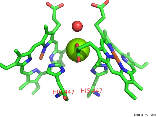
Mono view
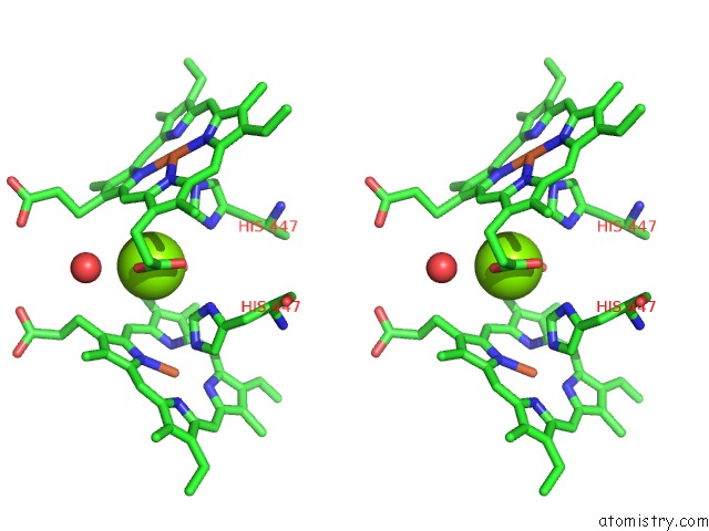
Stereo pair view
Mono view
Stereo pair view
A full contact list of Magnesium with other atoms in the Mg binding
site number 1 of Crystal Structure of Outer Cell Wall Cytochrome Ocwa within 5.0Å range:
|
Reference:
N.L.Costa,
B.Hermann,
V.Fourmond,
M.M.Faustino,
M.Teixeira,
O.Einsle,
C.M.Paquete,
R.O.Louro.
How Thermophilic Gram-Positive Organisms Perform Extracellular Electron Transfer: Characterization of the Cell Surface Terminal Reductase Ocwa. Mbio V. 10 2019.
ISSN: ESSN 2150-7511
PubMed: 31431546
DOI: 10.1128/MBIO.01210-19
Page generated: Tue Oct 1 03:00:00 2024
ISSN: ESSN 2150-7511
PubMed: 31431546
DOI: 10.1128/MBIO.01210-19
Last articles
Zn in 9J0NZn in 9J0O
Zn in 9J0P
Zn in 9FJX
Zn in 9EKB
Zn in 9C0F
Zn in 9CAH
Zn in 9CH0
Zn in 9CH3
Zn in 9CH1