Magnesium »
PDB 6iae-6ijj »
6ihl »
Magnesium in PDB 6ihl: Crystal Structure of Bacterial Serine Phosphatase
Enzymatic activity of Crystal Structure of Bacterial Serine Phosphatase
All present enzymatic activity of Crystal Structure of Bacterial Serine Phosphatase:
3.1.3.16;
3.1.3.16;
Protein crystallography data
The structure of Crystal Structure of Bacterial Serine Phosphatase, PDB code: 6ihl
was solved by
C.-G.Yang,
T.Yang,
with X-Ray Crystallography technique. A brief refinement statistics is given in the table below:
| Resolution Low / High (Å) | 45.74 / 1.57 |
| Space group | P 1 21 1 |
| Cell size a, b, c (Å), α, β, γ (°) | 46.669, 38.160, 64.891, 90.00, 101.47, 90.00 |
| R / Rfree (%) | 17 / 20.5 |
Magnesium Binding Sites:
The binding sites of Magnesium atom in the Crystal Structure of Bacterial Serine Phosphatase
(pdb code 6ihl). This binding sites where shown within
5.0 Angstroms radius around Magnesium atom.
In total 5 binding sites of Magnesium where determined in the Crystal Structure of Bacterial Serine Phosphatase, PDB code: 6ihl:
Jump to Magnesium binding site number: 1; 2; 3; 4; 5;
In total 5 binding sites of Magnesium where determined in the Crystal Structure of Bacterial Serine Phosphatase, PDB code: 6ihl:
Jump to Magnesium binding site number: 1; 2; 3; 4; 5;
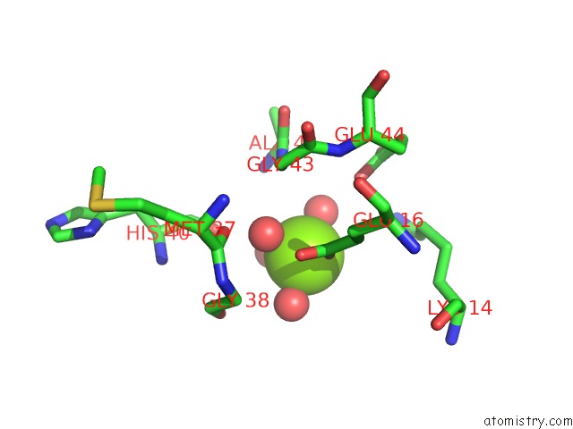
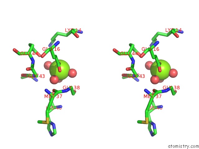
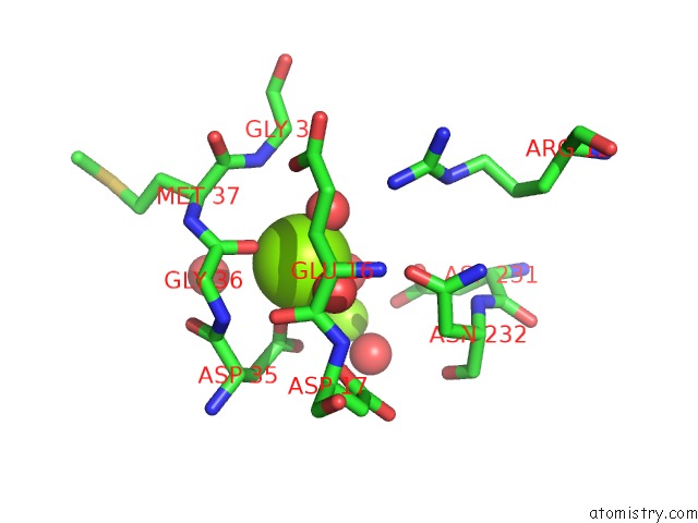
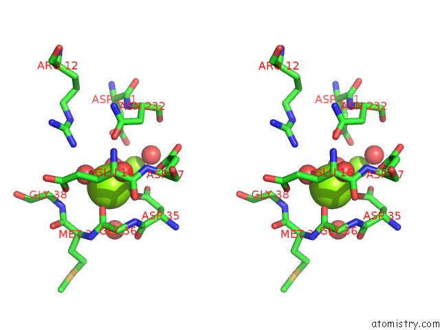
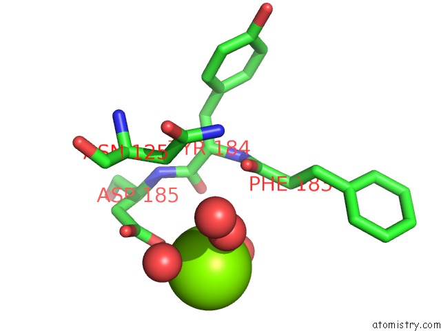
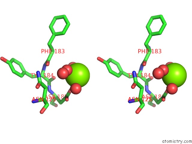
Magnesium binding site 1 out of 5 in 6ihl
Go back to
Magnesium binding site 1 out
of 5 in the Crystal Structure of Bacterial Serine Phosphatase
Mono view
Stereo pair view
Mono view
Stereo pair view
A full contact list of Magnesium with other atoms in the Mg binding
site number 1 of Crystal Structure of Bacterial Serine Phosphatase within 5.0Å range:
|
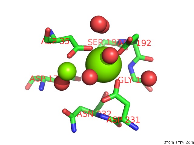
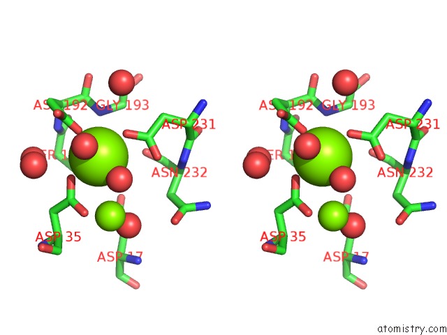
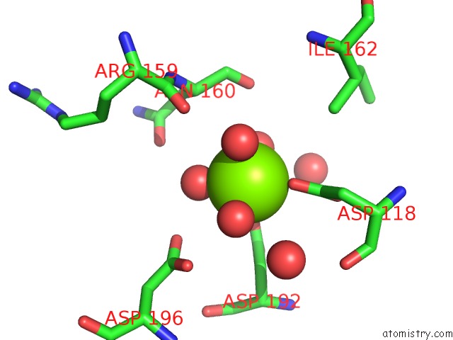
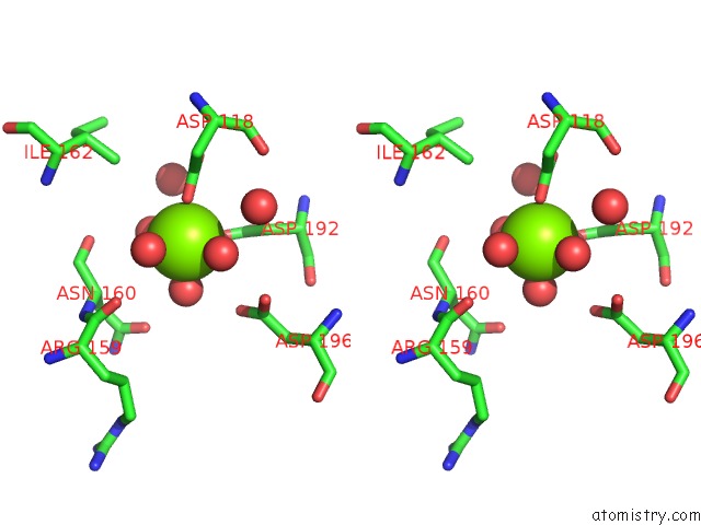
Magnesium binding site 2 out of 5 in 6ihl
Go back to
Magnesium binding site 2 out
of 5 in the Crystal Structure of Bacterial Serine Phosphatase
Mono view
Stereo pair view
Mono view
Stereo pair view
A full contact list of Magnesium with other atoms in the Mg binding
site number 2 of Crystal Structure of Bacterial Serine Phosphatase within 5.0Å range:
|
Magnesium binding site 3 out of 5 in 6ihl
Go back to
Magnesium binding site 3 out
of 5 in the Crystal Structure of Bacterial Serine Phosphatase
Mono view
Stereo pair view
Mono view
Stereo pair view
A full contact list of Magnesium with other atoms in the Mg binding
site number 3 of Crystal Structure of Bacterial Serine Phosphatase within 5.0Å range:
|
Magnesium binding site 4 out of 5 in 6ihl
Go back to
Magnesium binding site 4 out
of 5 in the Crystal Structure of Bacterial Serine Phosphatase
Mono view
Stereo pair view
Mono view
Stereo pair view
A full contact list of Magnesium with other atoms in the Mg binding
site number 4 of Crystal Structure of Bacterial Serine Phosphatase within 5.0Å range:
|
Magnesium binding site 5 out of 5 in 6ihl
Go back to
Magnesium binding site 5 out
of 5 in the Crystal Structure of Bacterial Serine Phosphatase
Mono view
Stereo pair view
Mono view
Stereo pair view
A full contact list of Magnesium with other atoms in the Mg binding
site number 5 of Crystal Structure of Bacterial Serine Phosphatase within 5.0Å range:
|
Reference:
T.Yang,
T.Liu,
J.Gan,
K.Yu,
K.Chen,
W.Xue,
L.Lan,
S.Yang,
C.G.Yang.
Structural Insight Into the Mechanism of Staphylococcus Aureus STP1 Phosphatase. Acs Infect Dis. V. 5 841 2019.
ISSN: ESSN 2373-8227
PubMed: 30868877
DOI: 10.1021/ACSINFECDIS.8B00316
Page generated: Tue Oct 1 03:22:18 2024
ISSN: ESSN 2373-8227
PubMed: 30868877
DOI: 10.1021/ACSINFECDIS.8B00316
Last articles
Zn in 9MJ5Zn in 9HNW
Zn in 9G0L
Zn in 9FNE
Zn in 9DZN
Zn in 9E0I
Zn in 9D32
Zn in 9DAK
Zn in 8ZXC
Zn in 8ZUF