Magnesium »
PDB 6ijj-6iug »
6im6 »
Magnesium in PDB 6im6: Crystal Structure of PDE4D Complexed with A Novel Inhibitor
Enzymatic activity of Crystal Structure of PDE4D Complexed with A Novel Inhibitor
All present enzymatic activity of Crystal Structure of PDE4D Complexed with A Novel Inhibitor:
3.1.4.53;
3.1.4.53;
Protein crystallography data
The structure of Crystal Structure of PDE4D Complexed with A Novel Inhibitor, PDB code: 6im6
was solved by
X.L.Zhang,
H.X.Su,
Y.C.Xu,
with X-Ray Crystallography technique. A brief refinement statistics is given in the table below:
| Resolution Low / High (Å) | 40.17 / 1.70 |
| Space group | P 21 21 21 |
| Cell size a, b, c (Å), α, β, γ (°) | 58.081, 80.346, 163.350, 90.00, 90.00, 90.00 |
| R / Rfree (%) | 21.1 / 24 |
Other elements in 6im6:
The structure of Crystal Structure of PDE4D Complexed with A Novel Inhibitor also contains other interesting chemical elements:
| Zinc | (Zn) | 2 atoms |
Magnesium Binding Sites:
The binding sites of Magnesium atom in the Crystal Structure of PDE4D Complexed with A Novel Inhibitor
(pdb code 6im6). This binding sites where shown within
5.0 Angstroms radius around Magnesium atom.
In total 4 binding sites of Magnesium where determined in the Crystal Structure of PDE4D Complexed with A Novel Inhibitor, PDB code: 6im6:
Jump to Magnesium binding site number: 1; 2; 3; 4;
In total 4 binding sites of Magnesium where determined in the Crystal Structure of PDE4D Complexed with A Novel Inhibitor, PDB code: 6im6:
Jump to Magnesium binding site number: 1; 2; 3; 4;
Magnesium binding site 1 out of 4 in 6im6
Go back to
Magnesium binding site 1 out
of 4 in the Crystal Structure of PDE4D Complexed with A Novel Inhibitor

Mono view
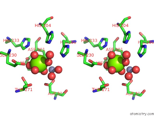
Stereo pair view
Mono view
Stereo pair view
A full contact list of Magnesium with other atoms in the Mg binding
site number 1 of Crystal Structure of PDE4D Complexed with A Novel Inhibitor within 5.0Å range:
|
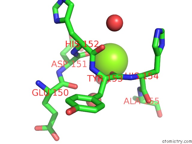
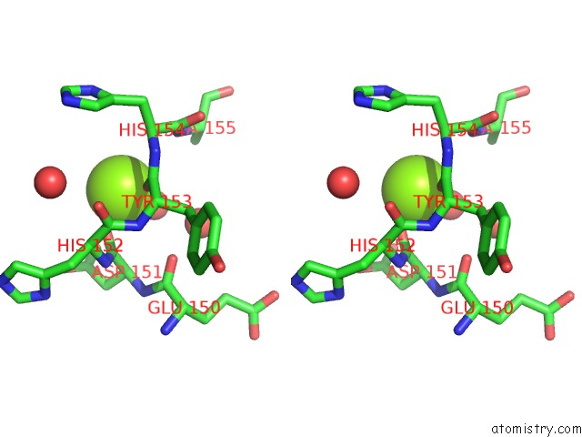
Magnesium binding site 2 out of 4 in 6im6
Go back to
Magnesium binding site 2 out
of 4 in the Crystal Structure of PDE4D Complexed with A Novel Inhibitor

Mono view

Stereo pair view
Mono view
Stereo pair view
A full contact list of Magnesium with other atoms in the Mg binding
site number 2 of Crystal Structure of PDE4D Complexed with A Novel Inhibitor within 5.0Å range:
|
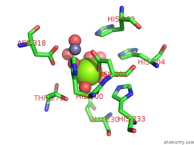
Magnesium binding site 3 out of 4 in 6im6
Go back to
Magnesium binding site 3 out
of 4 in the Crystal Structure of PDE4D Complexed with A Novel Inhibitor
Mono view
Stereo pair view
Mono view
Stereo pair view
A full contact list of Magnesium with other atoms in the Mg binding
site number 3 of Crystal Structure of PDE4D Complexed with A Novel Inhibitor within 5.0Å range:
|
Magnesium binding site 4 out of 4 in 6im6
Go back to
Magnesium binding site 4 out
of 4 in the Crystal Structure of PDE4D Complexed with A Novel Inhibitor
Mono view

Stereo pair view
Mono view
Stereo pair view
A full contact list of Magnesium with other atoms in the Mg binding
site number 4 of Crystal Structure of PDE4D Complexed with A Novel Inhibitor within 5.0Å range:
|
Reference:
X.Zhang,
G.Dong,
H.Li,
W.Chen,
J.Li,
C.Feng,
Z.Gu,
F.Zhu,
R.Zhang,
M.Li,
W.Tang,
H.Liu,
Y.Xu.
Structure-Aided Identification and Optimization of Tetrahydro-Isoquinolines As Novel PDE4 Inhibitors Leading to Discovery of An Effective Antipsoriasis Agent. J.Med.Chem. V. 62 5579 2019.
ISSN: ISSN 0022-2623
PubMed: 31099559
DOI: 10.1021/ACS.JMEDCHEM.9B00518
Page generated: Tue Oct 1 03:48:12 2024
ISSN: ISSN 0022-2623
PubMed: 31099559
DOI: 10.1021/ACS.JMEDCHEM.9B00518
Last articles
Zn in 9J0NZn in 9J0O
Zn in 9J0P
Zn in 9FJX
Zn in 9EKB
Zn in 9C0F
Zn in 9CAH
Zn in 9CH0
Zn in 9CH3
Zn in 9CH1