Magnesium »
PDB 6jwt-6k77 »
6jxm »
Magnesium in PDB 6jxm: Crystal Structure of PHI29 Prna Domain II
Protein crystallography data
The structure of Crystal Structure of PHI29 Prna Domain II, PDB code: 6jxm
was solved by
R.Cai,
I.R.Price,
F.Ding,
F.Wu,
T.Chen,
Y.Zhang,
G.Liu,
P.J.Jardine,
C.Lu,
A.Ke,
with X-Ray Crystallography technique. A brief refinement statistics is given in the table below:
| Resolution Low / High (Å) | 101.18 / 3.32 |
| Space group | I 4 2 2 |
| Cell size a, b, c (Å), α, β, γ (°) | 120.178, 120.178, 187.494, 90.00, 90.00, 90.00 |
| R / Rfree (%) | 22.3 / 26.5 |
Other elements in 6jxm:
The structure of Crystal Structure of PHI29 Prna Domain II also contains other interesting chemical elements:
| Barium | (Ba) | 15 atoms |
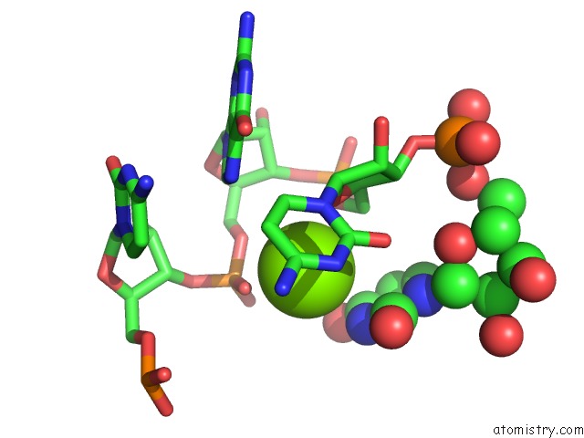
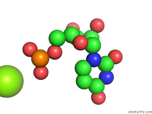
Magnesium Binding Sites:
The binding sites of Magnesium atom in the Crystal Structure of PHI29 Prna Domain II
(pdb code 6jxm). This binding sites where shown within
5.0 Angstroms radius around Magnesium atom.
In total 7 binding sites of Magnesium where determined in the Crystal Structure of PHI29 Prna Domain II, PDB code: 6jxm:
Jump to Magnesium binding site number: 1; 2; 3; 4; 5; 6; 7;
In total 7 binding sites of Magnesium where determined in the Crystal Structure of PHI29 Prna Domain II, PDB code: 6jxm:
Jump to Magnesium binding site number: 1; 2; 3; 4; 5; 6; 7;
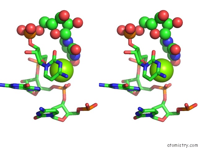
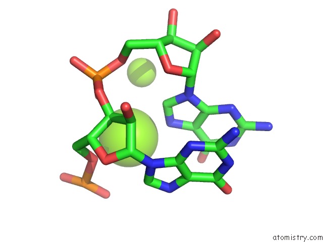
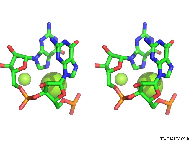
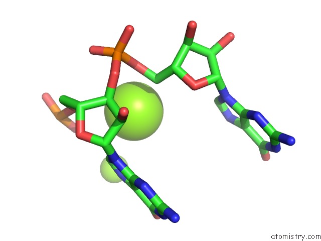
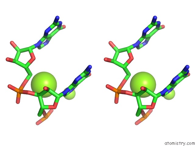
Magnesium binding site 1 out of 7 in 6jxm
Go back to
Magnesium binding site 1 out
of 7 in the Crystal Structure of PHI29 Prna Domain II
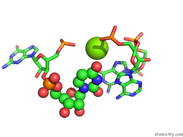
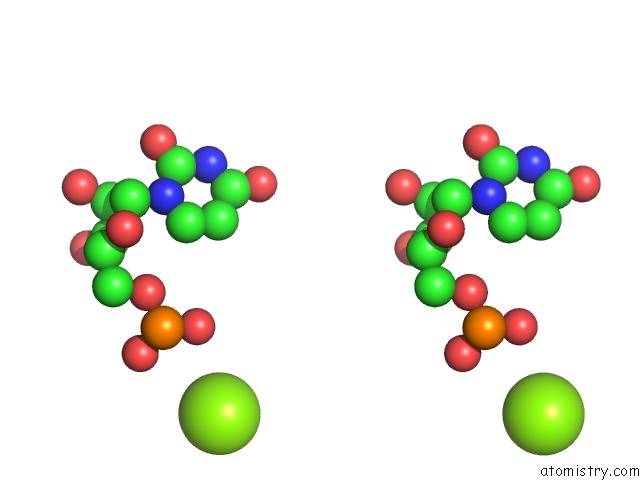
Mono view
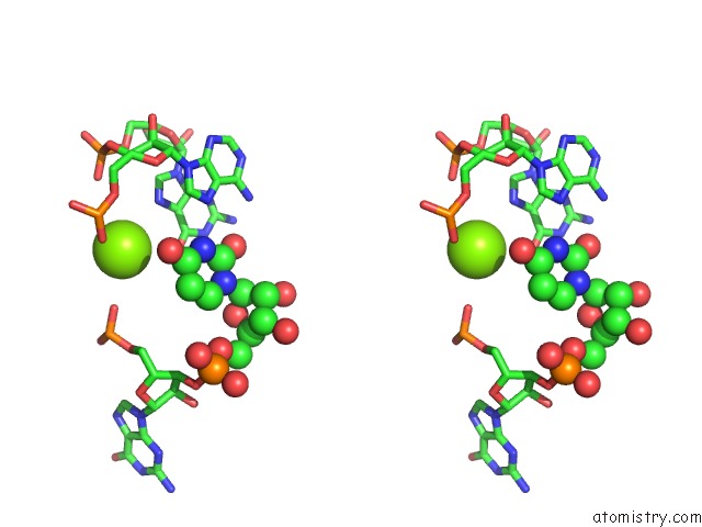
Stereo pair view
Mono view
Stereo pair view
A full contact list of Magnesium with other atoms in the Mg binding
site number 1 of Crystal Structure of PHI29 Prna Domain II within 5.0Å range:
|
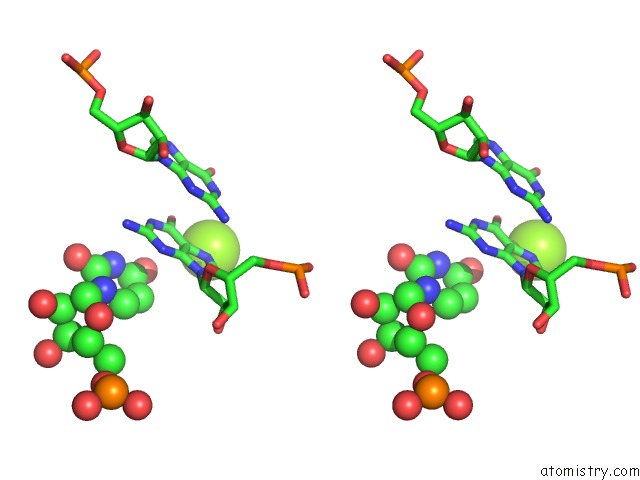
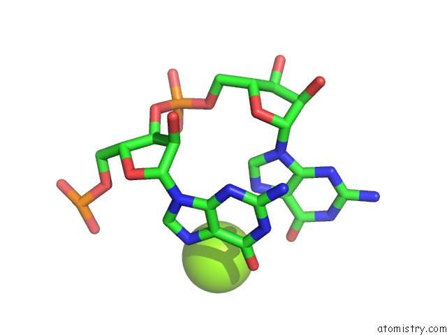
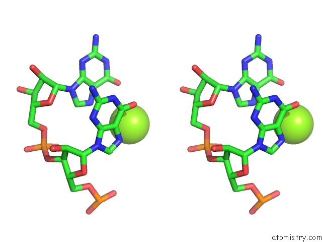
Magnesium binding site 2 out of 7 in 6jxm
Go back to
Magnesium binding site 2 out
of 7 in the Crystal Structure of PHI29 Prna Domain II
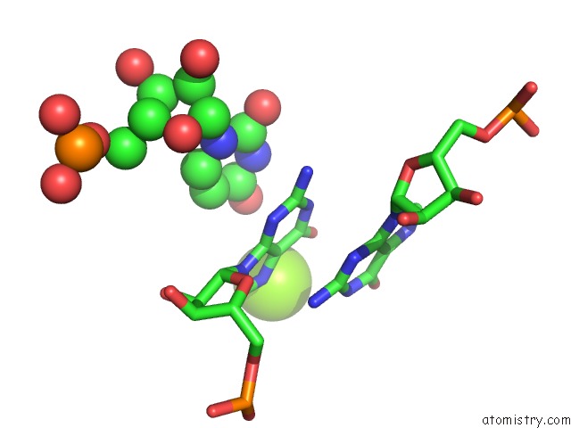
Mono view
Stereo pair view
Mono view
Stereo pair view
A full contact list of Magnesium with other atoms in the Mg binding
site number 2 of Crystal Structure of PHI29 Prna Domain II within 5.0Å range:
|
Magnesium binding site 3 out of 7 in 6jxm
Go back to
Magnesium binding site 3 out
of 7 in the Crystal Structure of PHI29 Prna Domain II
Mono view
Stereo pair view
Mono view
Stereo pair view
A full contact list of Magnesium with other atoms in the Mg binding
site number 3 of Crystal Structure of PHI29 Prna Domain II within 5.0Å range:
|
Magnesium binding site 4 out of 7 in 6jxm
Go back to
Magnesium binding site 4 out
of 7 in the Crystal Structure of PHI29 Prna Domain II
Mono view
Stereo pair view
Mono view
Stereo pair view
A full contact list of Magnesium with other atoms in the Mg binding
site number 4 of Crystal Structure of PHI29 Prna Domain II within 5.0Å range:
|
Magnesium binding site 5 out of 7 in 6jxm
Go back to
Magnesium binding site 5 out
of 7 in the Crystal Structure of PHI29 Prna Domain II
Mono view
Stereo pair view
Mono view
Stereo pair view
A full contact list of Magnesium with other atoms in the Mg binding
site number 5 of Crystal Structure of PHI29 Prna Domain II within 5.0Å range:
|
Magnesium binding site 6 out of 7 in 6jxm
Go back to
Magnesium binding site 6 out
of 7 in the Crystal Structure of PHI29 Prna Domain II
Mono view
Stereo pair view
Mono view
Stereo pair view
A full contact list of Magnesium with other atoms in the Mg binding
site number 6 of Crystal Structure of PHI29 Prna Domain II within 5.0Å range:
|
Magnesium binding site 7 out of 7 in 6jxm
Go back to
Magnesium binding site 7 out
of 7 in the Crystal Structure of PHI29 Prna Domain II
Mono view
Stereo pair view
Mono view
Stereo pair view
A full contact list of Magnesium with other atoms in the Mg binding
site number 7 of Crystal Structure of PHI29 Prna Domain II within 5.0Å range:
|
Reference:
R.Cai,
I.R.Price,
F.Ding,
F.Wu,
T.Chen,
Y.Zhang,
G.Liu,
P.J.Jardine,
C.Lu,
A.Ke.
Atp/Adp Modulates GP16-Prna Conformational Change in the PHI29 Dna Packaging Motor. Nucleic Acids Res. V. 47 9818 2019.
ISSN: ESSN 1362-4962
PubMed: 31396619
DOI: 10.1093/NAR/GKZ692
Page generated: Tue Oct 1 06:20:53 2024
ISSN: ESSN 1362-4962
PubMed: 31396619
DOI: 10.1093/NAR/GKZ692
Last articles
Zn in 9MJ5Zn in 9HNW
Zn in 9G0L
Zn in 9FNE
Zn in 9DZN
Zn in 9E0I
Zn in 9D32
Zn in 9DAK
Zn in 8ZXC
Zn in 8ZUF