Magnesium »
PDB 6jws-6k74 »
6k0h »
Magnesium in PDB 6k0h: Crystal Structure of Udp-Glucose 4-Epimerase From Bifidobacterium Longum in Complex with Nad+ and Udp-Glcnac
Enzymatic activity of Crystal Structure of Udp-Glucose 4-Epimerase From Bifidobacterium Longum in Complex with Nad+ and Udp-Glcnac
All present enzymatic activity of Crystal Structure of Udp-Glucose 4-Epimerase From Bifidobacterium Longum in Complex with Nad+ and Udp-Glcnac:
5.1.3.2;
5.1.3.2;
Protein crystallography data
The structure of Crystal Structure of Udp-Glucose 4-Epimerase From Bifidobacterium Longum in Complex with Nad+ and Udp-Glcnac, PDB code: 6k0h
was solved by
Y.-W.Nam,
M.Nishimoto,
T.Arakawa,
M.Kitaoka,
S.Fushinobu,
with X-Ray Crystallography technique. A brief refinement statistics is given in the table below:
| Resolution Low / High (Å) | 34.75 / 2.00 |
| Space group | P 65 2 2 |
| Cell size a, b, c (Å), α, β, γ (°) | 69.450, 69.450, 321.986, 90.00, 90.00, 120.00 |
| R / Rfree (%) | 18.1 / 22.1 |
Magnesium Binding Sites:
The binding sites of Magnesium atom in the Crystal Structure of Udp-Glucose 4-Epimerase From Bifidobacterium Longum in Complex with Nad+ and Udp-Glcnac
(pdb code 6k0h). This binding sites where shown within
5.0 Angstroms radius around Magnesium atom.
In total 2 binding sites of Magnesium where determined in the Crystal Structure of Udp-Glucose 4-Epimerase From Bifidobacterium Longum in Complex with Nad+ and Udp-Glcnac, PDB code: 6k0h:
Jump to Magnesium binding site number: 1; 2;
In total 2 binding sites of Magnesium where determined in the Crystal Structure of Udp-Glucose 4-Epimerase From Bifidobacterium Longum in Complex with Nad+ and Udp-Glcnac, PDB code: 6k0h:
Jump to Magnesium binding site number: 1; 2;
Magnesium binding site 1 out of 2 in 6k0h
Go back to
Magnesium binding site 1 out
of 2 in the Crystal Structure of Udp-Glucose 4-Epimerase From Bifidobacterium Longum in Complex with Nad+ and Udp-Glcnac
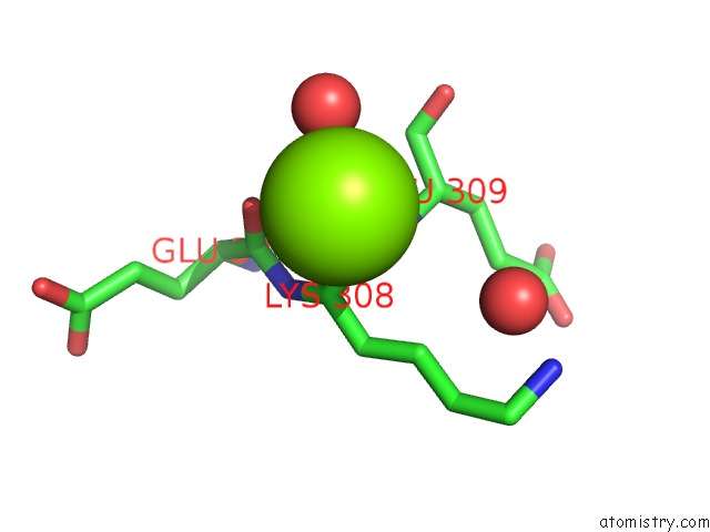
Mono view
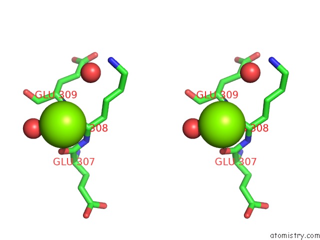
Stereo pair view
Mono view
Stereo pair view
A full contact list of Magnesium with other atoms in the Mg binding
site number 1 of Crystal Structure of Udp-Glucose 4-Epimerase From Bifidobacterium Longum in Complex with Nad+ and Udp-Glcnac within 5.0Å range:
|
Magnesium binding site 2 out of 2 in 6k0h
Go back to
Magnesium binding site 2 out
of 2 in the Crystal Structure of Udp-Glucose 4-Epimerase From Bifidobacterium Longum in Complex with Nad+ and Udp-Glcnac
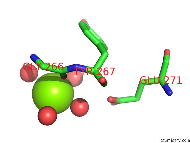
Mono view
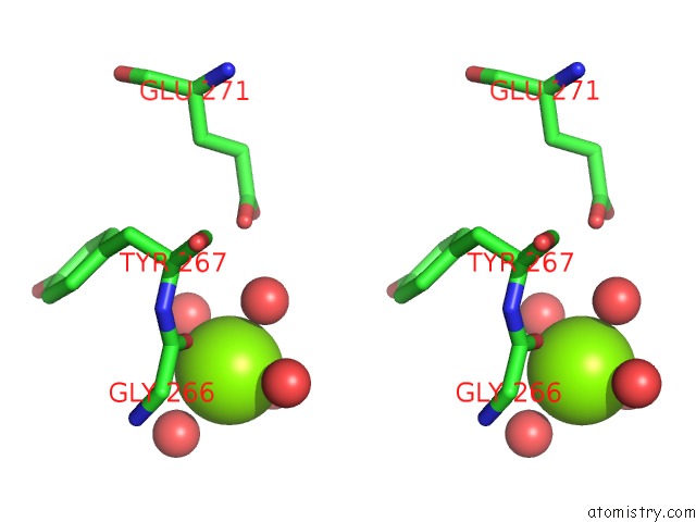
Stereo pair view
Mono view
Stereo pair view
A full contact list of Magnesium with other atoms in the Mg binding
site number 2 of Crystal Structure of Udp-Glucose 4-Epimerase From Bifidobacterium Longum in Complex with Nad+ and Udp-Glcnac within 5.0Å range:
|
Reference:
Y.W.Nam,
M.Nishimoto,
T.Arakawa,
M.Kitaoka,
S.Fushinobu.
Structural Basis For Broad Substrate Specificity of Udp-Glucose 4-Epimerase in the Human Milk Oligosaccharide Catabolic Pathway of Bifidobacterium Longum. Sci Rep V. 9 11081 2019.
ISSN: ESSN 2045-2322
PubMed: 31366978
DOI: 10.1038/S41598-019-47591-W
Page generated: Tue Oct 1 06:21:44 2024
ISSN: ESSN 2045-2322
PubMed: 31366978
DOI: 10.1038/S41598-019-47591-W
Last articles
Zn in 9J0NZn in 9J0O
Zn in 9J0P
Zn in 9FJX
Zn in 9EKB
Zn in 9C0F
Zn in 9CAH
Zn in 9CH0
Zn in 9CH3
Zn in 9CH1