Magnesium »
PDB 6kqm-6l59 »
6kz6 »
Magnesium in PDB 6kz6: Crystal Structure of Asfv Dutpase
Protein crystallography data
The structure of Crystal Structure of Asfv Dutpase, PDB code: 6kz6
was solved by
Y.Guo,
C.Chen,
G.B.Li,
L.Cao,
C.W.Wang,
with X-Ray Crystallography technique. A brief refinement statistics is given in the table below:
| Resolution Low / High (Å) | 33.13 / 2.19 |
| Space group | P 21 21 21 |
| Cell size a, b, c (Å), α, β, γ (°) | 66.256, 67.278, 116.912, 90.00, 90.00, 90.00 |
| R / Rfree (%) | 19.6 / 22.8 |
Magnesium Binding Sites:
The binding sites of Magnesium atom in the Crystal Structure of Asfv Dutpase
(pdb code 6kz6). This binding sites where shown within
5.0 Angstroms radius around Magnesium atom.
In total 2 binding sites of Magnesium where determined in the Crystal Structure of Asfv Dutpase, PDB code: 6kz6:
Jump to Magnesium binding site number: 1; 2;
In total 2 binding sites of Magnesium where determined in the Crystal Structure of Asfv Dutpase, PDB code: 6kz6:
Jump to Magnesium binding site number: 1; 2;
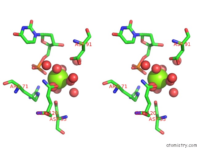
Magnesium binding site 1 out of 2 in 6kz6
Go back to
Magnesium binding site 1 out
of 2 in the Crystal Structure of Asfv Dutpase

Mono view

Stereo pair view
Mono view
Stereo pair view
A full contact list of Magnesium with other atoms in the Mg binding
site number 1 of Crystal Structure of Asfv Dutpase within 5.0Å range:
|
Magnesium binding site 2 out of 2 in 6kz6
Go back to
Magnesium binding site 2 out
of 2 in the Crystal Structure of Asfv Dutpase

Mono view
Stereo pair view
Mono view
Stereo pair view
A full contact list of Magnesium with other atoms in the Mg binding
site number 2 of Crystal Structure of Asfv Dutpase within 5.0Å range:
|
Reference:
G.B.Li,
C.W.Wang,
M.Yang,
L.Cao,
D.Fu,
X.Liu,
D.Sun,
C.Chen,
Y.Wang,
Z.Jia,
C.Yang,
Y.Guo,
Z.Rao.
Structural Insight Into the African Swine Fever Virus Dutpase Reveals A Novel Folding Pattern in the Dutpase Family. J.Virol. 2019.
ISSN: ESSN 1098-5514
PubMed: 31748385
DOI: 10.1128/JVI.01698-19
Page generated: Tue Oct 1 10:01:01 2024
ISSN: ESSN 1098-5514
PubMed: 31748385
DOI: 10.1128/JVI.01698-19
Last articles
F in 7NVVF in 7NVO
F in 7NTH
F in 7NTI
F in 7NPC
F in 7NRG
F in 7NR5
F in 7NQS
F in 7NOS
F in 7NP5