Magnesium »
PDB 6l5l-6ll1 »
6l8s »
Magnesium in PDB 6l8s: High Resolution Crystal Structure of Crustacean Hemocyanin.
Protein crystallography data
The structure of High Resolution Crystal Structure of Crustacean Hemocyanin., PDB code: 6l8s
was solved by
T.Masuda,
B.Mikami,
S.Baba,
with X-Ray Crystallography technique. A brief refinement statistics is given in the table below:
| Resolution Low / High (Å) | 42.70 / 1.58 |
| Space group | C 2 2 21 |
| Cell size a, b, c (Å), α, β, γ (°) | 119.454, 207.596, 187.364, 90.00, 90.00, 90.00 |
| R / Rfree (%) | 14.9 / 18.6 |
Other elements in 6l8s:
The structure of High Resolution Crystal Structure of Crustacean Hemocyanin. also contains other interesting chemical elements:
| Copper | (Cu) | 6 atoms |
| Chlorine | (Cl) | 1 atom |
Magnesium Binding Sites:
The binding sites of Magnesium atom in the High Resolution Crystal Structure of Crustacean Hemocyanin.
(pdb code 6l8s). This binding sites where shown within
5.0 Angstroms radius around Magnesium atom.
In total 5 binding sites of Magnesium where determined in the High Resolution Crystal Structure of Crustacean Hemocyanin., PDB code: 6l8s:
Jump to Magnesium binding site number: 1; 2; 3; 4; 5;
In total 5 binding sites of Magnesium where determined in the High Resolution Crystal Structure of Crustacean Hemocyanin., PDB code: 6l8s:
Jump to Magnesium binding site number: 1; 2; 3; 4; 5;
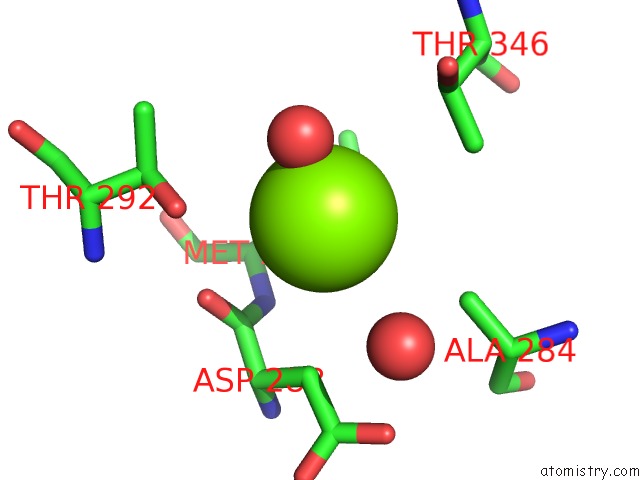
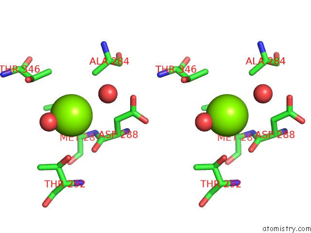
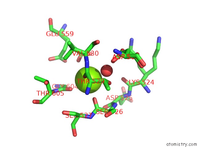
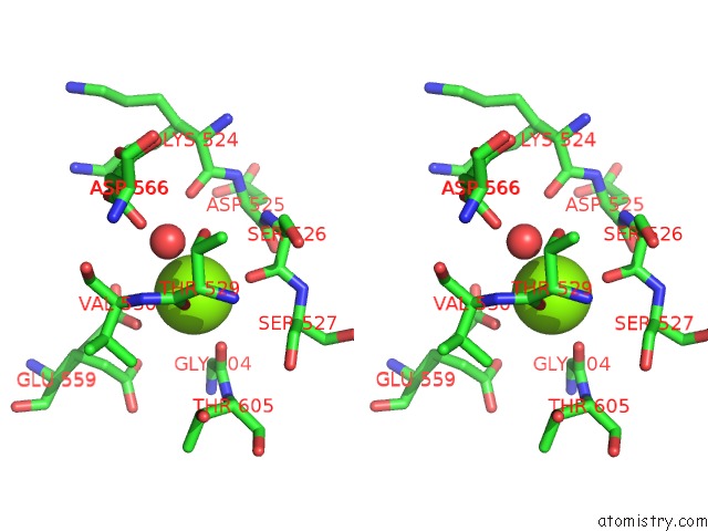
Magnesium binding site 1 out of 5 in 6l8s
Go back to
Magnesium binding site 1 out
of 5 in the High Resolution Crystal Structure of Crustacean Hemocyanin.
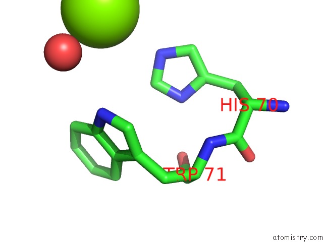
Mono view
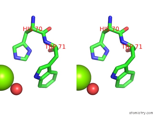
Stereo pair view
Mono view
Stereo pair view
A full contact list of Magnesium with other atoms in the Mg binding
site number 1 of High Resolution Crystal Structure of Crustacean Hemocyanin. within 5.0Å range:
|
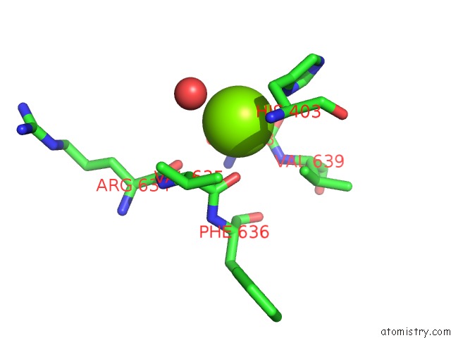
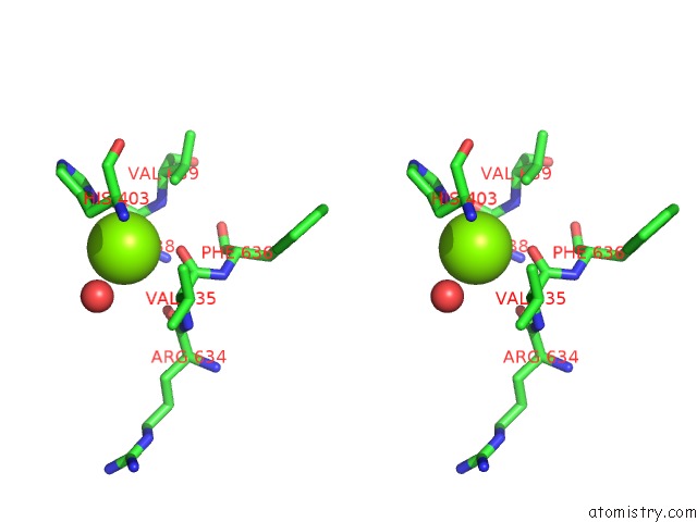
Magnesium binding site 2 out of 5 in 6l8s
Go back to
Magnesium binding site 2 out
of 5 in the High Resolution Crystal Structure of Crustacean Hemocyanin.
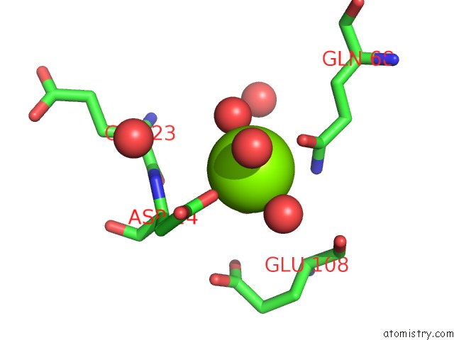
Mono view
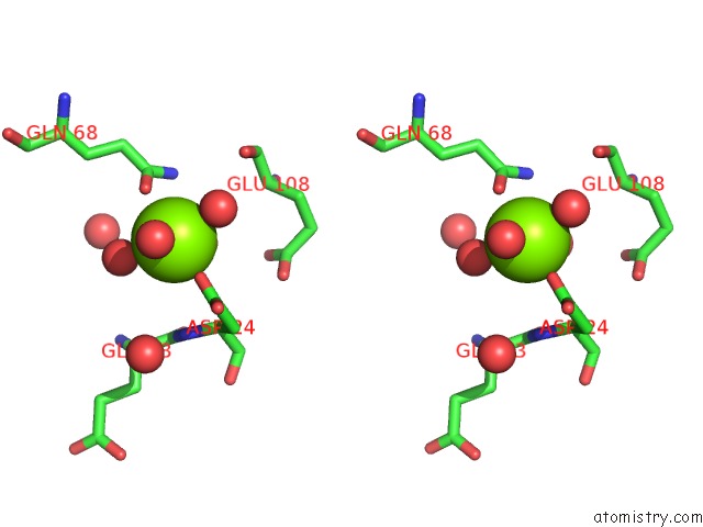
Stereo pair view
Mono view
Stereo pair view
A full contact list of Magnesium with other atoms in the Mg binding
site number 2 of High Resolution Crystal Structure of Crustacean Hemocyanin. within 5.0Å range:
|
Magnesium binding site 3 out of 5 in 6l8s
Go back to
Magnesium binding site 3 out
of 5 in the High Resolution Crystal Structure of Crustacean Hemocyanin.
Mono view
Stereo pair view
Mono view
Stereo pair view
A full contact list of Magnesium with other atoms in the Mg binding
site number 3 of High Resolution Crystal Structure of Crustacean Hemocyanin. within 5.0Å range:
|
Magnesium binding site 4 out of 5 in 6l8s
Go back to
Magnesium binding site 4 out
of 5 in the High Resolution Crystal Structure of Crustacean Hemocyanin.
Mono view
Stereo pair view
Mono view
Stereo pair view
A full contact list of Magnesium with other atoms in the Mg binding
site number 4 of High Resolution Crystal Structure of Crustacean Hemocyanin. within 5.0Å range:
|
Magnesium binding site 5 out of 5 in 6l8s
Go back to
Magnesium binding site 5 out
of 5 in the High Resolution Crystal Structure of Crustacean Hemocyanin.
Mono view
Stereo pair view
Mono view
Stereo pair view
A full contact list of Magnesium with other atoms in the Mg binding
site number 5 of High Resolution Crystal Structure of Crustacean Hemocyanin. within 5.0Å range:
|
Reference:
T.Masuda,
S.Baba,
K.Matsuo,
S.Ito,
B.Mikami.
The High-Resolution Crystal Structure of Lobster Hemocyanin Shows Its Enzymatic Capability As A Phenoloxidase. Arch.Biochem.Biophys. 08370 2020.
ISSN: ESSN 1096-0384
PubMed: 32380017
DOI: 10.1016/J.ABB.2020.108370
Page generated: Tue Oct 1 10:22:25 2024
ISSN: ESSN 1096-0384
PubMed: 32380017
DOI: 10.1016/J.ABB.2020.108370
Last articles
Cl in 7SINCl in 7SII
Cl in 7SIL
Cl in 7SHV
Cl in 7SHG
Cl in 7SIE
Cl in 7SGV
Cl in 7SGY
Cl in 7SGX
Cl in 7SGW