Magnesium »
PDB 6l5o-6ll4 »
6ljj »
Magnesium in PDB 6ljj: Swine Dutpase in Complex with Alpha,Beta-Iminodutp and Magnesium Ion
Protein crystallography data
The structure of Swine Dutpase in Complex with Alpha,Beta-Iminodutp and Magnesium Ion, PDB code: 6ljj
was solved by
R.Liang,
G.Q.Peng,
with X-Ray Crystallography technique. A brief refinement statistics is given in the table below:
| Resolution Low / High (Å) | 27.46 / 1.89 |
| Space group | F 41 3 2 |
| Cell size a, b, c (Å), α, β, γ (°) | 162.386, 162.386, 162.386, 90.00, 90.00, 90.00 |
| R / Rfree (%) | 19.7 / 23.1 |
Magnesium Binding Sites:
The binding sites of Magnesium atom in the Swine Dutpase in Complex with Alpha,Beta-Iminodutp and Magnesium Ion
(pdb code 6ljj). This binding sites where shown within
5.0 Angstroms radius around Magnesium atom.
In total only one binding site of Magnesium was determined in the Swine Dutpase in Complex with Alpha,Beta-Iminodutp and Magnesium Ion, PDB code: 6ljj:
In total only one binding site of Magnesium was determined in the Swine Dutpase in Complex with Alpha,Beta-Iminodutp and Magnesium Ion, PDB code: 6ljj:
Magnesium binding site 1 out of 1 in 6ljj
Go back to
Magnesium binding site 1 out
of 1 in the Swine Dutpase in Complex with Alpha,Beta-Iminodutp and Magnesium Ion
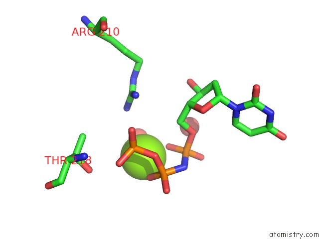
Mono view
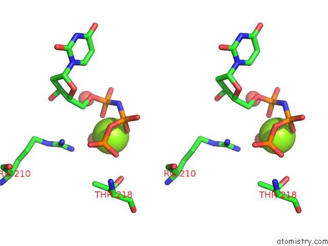
Stereo pair view
Mono view
Stereo pair view
A full contact list of Magnesium with other atoms in the Mg binding
site number 1 of Swine Dutpase in Complex with Alpha,Beta-Iminodutp and Magnesium Ion within 5.0Å range:
|
Reference:
R.Liang,
G.Wang,
D.Zhang,
G.Ye,
M.Li,
Y.Shi,
J.Shi,
H.Chen,
G.Peng.
Structural Comparisons of Host and African Swine Fever Virus Dutpases Reveal New Clues For Inhibitor Development. J.Biol.Chem. 2020.
ISSN: ESSN 1083-351X
PubMed: 33139328
DOI: 10.1074/JBC.RA120.014005
Page generated: Wed Aug 13 11:22:38 2025
ISSN: ESSN 1083-351X
PubMed: 33139328
DOI: 10.1074/JBC.RA120.014005
Last articles
Mg in 6YARMg in 6YAL
Mg in 6YA8
Mg in 6Y64
Mg in 6Y8D
Mg in 6YA5
Mg in 6Y8E
Mg in 6Y8B
Mg in 6Y8C
Mg in 6Y8A