Magnesium »
PDB 6ll8-6ly7 »
6lm1 »
Magnesium in PDB 6lm1: The Crystal Structure of Cyanorhodopsin (Cyr) N4075R From Cyanobacteria Tolypothrix Sp. Nies-4075
Protein crystallography data
The structure of The Crystal Structure of Cyanorhodopsin (Cyr) N4075R From Cyanobacteria Tolypothrix Sp. Nies-4075, PDB code: 6lm1
was solved by
T.Hosaka,
T.Kimura-Someya,
M.Shirouzu,
with X-Ray Crystallography technique. A brief refinement statistics is given in the table below:
| Resolution Low / High (Å) | 45.48 / 1.90 |
| Space group | C 2 2 21 |
| Cell size a, b, c (Å), α, β, γ (°) | 107.920, 129.666, 127.626, 90.00, 90.00, 90.00 |
| R / Rfree (%) | 16.5 / 20.4 |
Magnesium Binding Sites:
The binding sites of Magnesium atom in the The Crystal Structure of Cyanorhodopsin (Cyr) N4075R From Cyanobacteria Tolypothrix Sp. Nies-4075
(pdb code 6lm1). This binding sites where shown within
5.0 Angstroms radius around Magnesium atom.
In total 3 binding sites of Magnesium where determined in the The Crystal Structure of Cyanorhodopsin (Cyr) N4075R From Cyanobacteria Tolypothrix Sp. Nies-4075, PDB code: 6lm1:
Jump to Magnesium binding site number: 1; 2; 3;
In total 3 binding sites of Magnesium where determined in the The Crystal Structure of Cyanorhodopsin (Cyr) N4075R From Cyanobacteria Tolypothrix Sp. Nies-4075, PDB code: 6lm1:
Jump to Magnesium binding site number: 1; 2; 3;
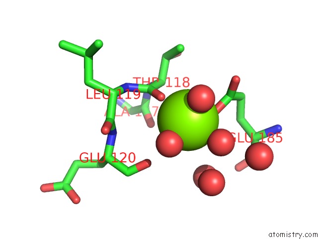
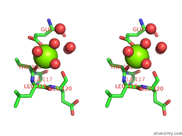
Magnesium binding site 1 out of 3 in 6lm1
Go back to
Magnesium binding site 1 out
of 3 in the The Crystal Structure of Cyanorhodopsin (Cyr) N4075R From Cyanobacteria Tolypothrix Sp. Nies-4075
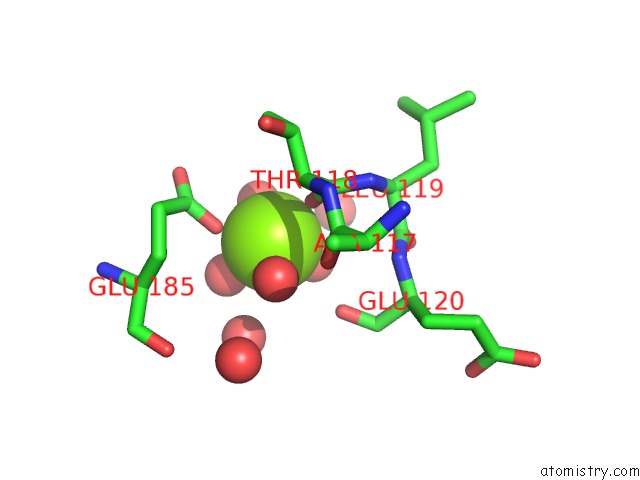
Mono view
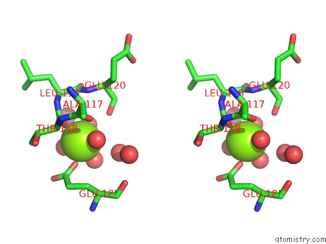
Stereo pair view
Mono view
Stereo pair view
A full contact list of Magnesium with other atoms in the Mg binding
site number 1 of The Crystal Structure of Cyanorhodopsin (Cyr) N4075R From Cyanobacteria Tolypothrix Sp. Nies-4075 within 5.0Å range:
|
Magnesium binding site 2 out of 3 in 6lm1
Go back to
Magnesium binding site 2 out
of 3 in the The Crystal Structure of Cyanorhodopsin (Cyr) N4075R From Cyanobacteria Tolypothrix Sp. Nies-4075
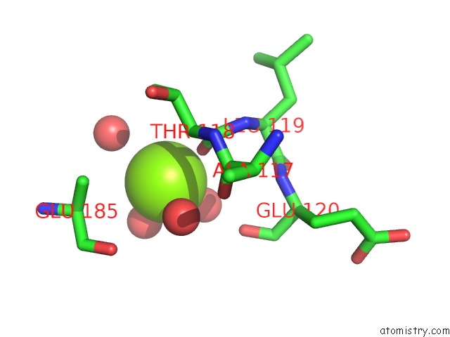
Mono view
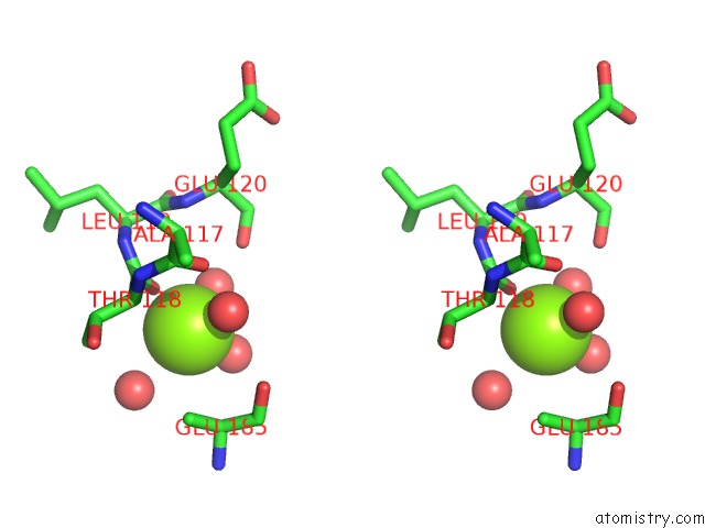
Stereo pair view
Mono view
Stereo pair view
A full contact list of Magnesium with other atoms in the Mg binding
site number 2 of The Crystal Structure of Cyanorhodopsin (Cyr) N4075R From Cyanobacteria Tolypothrix Sp. Nies-4075 within 5.0Å range:
|
Magnesium binding site 3 out of 3 in 6lm1
Go back to
Magnesium binding site 3 out
of 3 in the The Crystal Structure of Cyanorhodopsin (Cyr) N4075R From Cyanobacteria Tolypothrix Sp. Nies-4075
Mono view
Stereo pair view
Mono view
Stereo pair view
A full contact list of Magnesium with other atoms in the Mg binding
site number 3 of The Crystal Structure of Cyanorhodopsin (Cyr) N4075R From Cyanobacteria Tolypothrix Sp. Nies-4075 within 5.0Å range:
|
Reference:
M.Hasegawa,
T.Hosaka,
K.Kojima,
Y.Nishimura,
Y.Nakajima,
T.Kimura-Someya,
M.Shirouzu,
Y.Sudo,
S.Yoshizawa.
A Unique Clade of Light-Driven Proton-Pumping Rhodopsins Evolved in the Cyanobacterial Lineage. Sci Rep V. 10 16752 2020.
ISSN: ESSN 2045-2322
PubMed: 33028840
DOI: 10.1038/S41598-020-73606-Y
Page generated: Wed Aug 13 11:24:23 2025
ISSN: ESSN 2045-2322
PubMed: 33028840
DOI: 10.1038/S41598-020-73606-Y
Last articles
Mg in 6QPHMg in 6QV0
Mg in 6QUZ
Mg in 6QUY
Mg in 6QUX
Mg in 6QUW
Mg in 6QUV
Mg in 6QUU
Mg in 6QUS
Mg in 6QTN