Magnesium »
PDB 6ll8-6ly7 »
6luh »
Magnesium in PDB 6luh: High Resolution Structure of N(Omega)-Hydroxy-L-Arginine Hydrolase
Enzymatic activity of High Resolution Structure of N(Omega)-Hydroxy-L-Arginine Hydrolase
All present enzymatic activity of High Resolution Structure of N(Omega)-Hydroxy-L-Arginine Hydrolase:
3.5.3.25;
3.5.3.25;
Protein crystallography data
The structure of High Resolution Structure of N(Omega)-Hydroxy-L-Arginine Hydrolase, PDB code: 6luh
was solved by
K.Oda,
Y.Matoba,
with X-Ray Crystallography technique. A brief refinement statistics is given in the table below:
| Resolution Low / High (Å) | 44.09 / 1.50 |
| Space group | P 1 |
| Cell size a, b, c (Å), α, β, γ (°) | 46.326, 47.057, 59.261, 83.57, 84.63, 70.13 |
| R / Rfree (%) | 20 / 23 |
Other elements in 6luh:
The structure of High Resolution Structure of N(Omega)-Hydroxy-L-Arginine Hydrolase also contains other interesting chemical elements:
| Manganese | (Mn) | 4 atoms |
Magnesium Binding Sites:
The binding sites of Magnesium atom in the High Resolution Structure of N(Omega)-Hydroxy-L-Arginine Hydrolase
(pdb code 6luh). This binding sites where shown within
5.0 Angstroms radius around Magnesium atom.
In total only one binding site of Magnesium was determined in the High Resolution Structure of N(Omega)-Hydroxy-L-Arginine Hydrolase, PDB code: 6luh:
In total only one binding site of Magnesium was determined in the High Resolution Structure of N(Omega)-Hydroxy-L-Arginine Hydrolase, PDB code: 6luh:
Magnesium binding site 1 out of 1 in 6luh
Go back to
Magnesium binding site 1 out
of 1 in the High Resolution Structure of N(Omega)-Hydroxy-L-Arginine Hydrolase
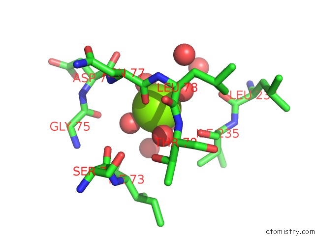
Mono view
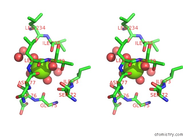
Stereo pair view
Mono view
Stereo pair view
A full contact list of Magnesium with other atoms in the Mg binding
site number 1 of High Resolution Structure of N(Omega)-Hydroxy-L-Arginine Hydrolase within 5.0Å range:
|
Reference:
K.Oda,
N.Shimotani,
T.Kuroda,
Y.Matoba.
Crystal Structure of An Nomega-Hydroxy-L-Arginine Hydrolase Found in the D-Cycloserine Biosynthetic Pathway. Acta Crystallogr D Struct V. 76 506 2020BIOL.
ISSN: ISSN 2059-7983
PubMed: 32496212
DOI: 10.1107/S2059798320004908
Page generated: Wed Aug 13 11:29:36 2025
ISSN: ISSN 2059-7983
PubMed: 32496212
DOI: 10.1107/S2059798320004908
Last articles
Mg in 6R29Mg in 6R1B
Mg in 6QXL
Mg in 6R1N
Mg in 6R10
Mg in 6R0Z
Mg in 6R0Y
Mg in 6R0W
Mg in 6QZY
Mg in 6QZK