Magnesium »
PDB 6lya-6m6a »
6m2y »
Magnesium in PDB 6m2y: Crystal Structure of A Formolase, Bfd Variant M6 From Pseudomonas Putida
Enzymatic activity of Crystal Structure of A Formolase, Bfd Variant M6 From Pseudomonas Putida
All present enzymatic activity of Crystal Structure of A Formolase, Bfd Variant M6 From Pseudomonas Putida:
4.1.1.7;
4.1.1.7;
Protein crystallography data
The structure of Crystal Structure of A Formolase, Bfd Variant M6 From Pseudomonas Putida, PDB code: 6m2y
was solved by
H.L.Wei,
W.D.Liu,
T.Z.Li,
L.L.Zhu,
with X-Ray Crystallography technique. A brief refinement statistics is given in the table below:
| Resolution Low / High (Å) | 24.60 / 2.10 |
| Space group | I 2 2 2 |
| Cell size a, b, c (Å), α, β, γ (°) | 103.515, 106.674, 116.548, 90, 90, 90 |
| R / Rfree (%) | 15.4 / 18.9 |
Magnesium Binding Sites:
The binding sites of Magnesium atom in the Crystal Structure of A Formolase, Bfd Variant M6 From Pseudomonas Putida
(pdb code 6m2y). This binding sites where shown within
5.0 Angstroms radius around Magnesium atom.
In total only one binding site of Magnesium was determined in the Crystal Structure of A Formolase, Bfd Variant M6 From Pseudomonas Putida, PDB code: 6m2y:
In total only one binding site of Magnesium was determined in the Crystal Structure of A Formolase, Bfd Variant M6 From Pseudomonas Putida, PDB code: 6m2y:
Magnesium binding site 1 out of 1 in 6m2y
Go back to
Magnesium binding site 1 out
of 1 in the Crystal Structure of A Formolase, Bfd Variant M6 From Pseudomonas Putida
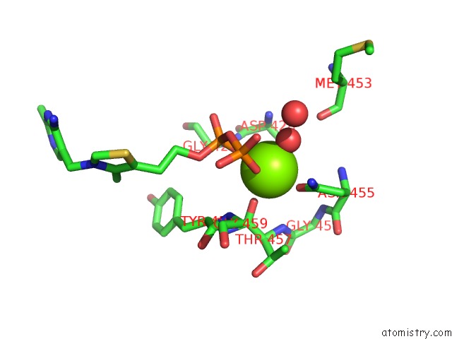
Mono view
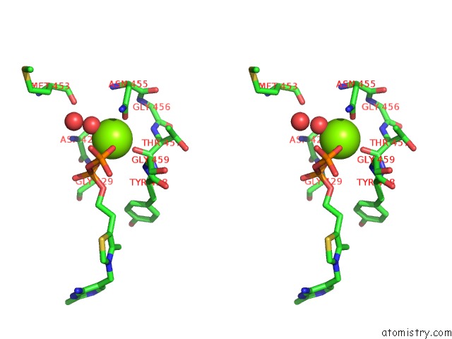
Stereo pair view
Mono view
Stereo pair view
A full contact list of Magnesium with other atoms in the Mg binding
site number 1 of Crystal Structure of A Formolase, Bfd Variant M6 From Pseudomonas Putida within 5.0Å range:
|
Reference:
T.Li,
Z.Tang,
H.Wei,
Z.Tan,
P.Liu,
J.Li,
Y.Zheng,
J.Lin,
W.Liu,
H.Jiang,
H.Liu,
L.Zhu,
Y.Ma.
Totally Atom-Economical Synthesis of Lactic Acid From Formaldehyde: Combined Bio-Carboligation and Chemo-Rearrangement Without the Isolation of Intermediate. Green Chem V. 22 6809 2020.
ISSN: ISSN 1463-9262
DOI: 10.1039/D0GC02433C
Page generated: Wed Aug 13 11:58:11 2025
ISSN: ISSN 1463-9262
DOI: 10.1039/D0GC02433C
Last articles
Mg in 7JGAMg in 7JG5
Mg in 7JFP
Mg in 7GYI
Mg in 7GYH
Mg in 7GYG
Mg in 7GYF
Mg in 7GYE
Mg in 7GYD
Mg in 7FSD