Magnesium »
PDB 6m60-6mlh »
6mgl »
Magnesium in PDB 6mgl: Crystal Structure of the Catalytic Domain From GH74 Enzyme POGH74 From Paenibacillus Odorifer, D60A Mutant in Complex with Xxlg and Xgxxlg Xyloglucan
Protein crystallography data
The structure of Crystal Structure of the Catalytic Domain From GH74 Enzyme POGH74 From Paenibacillus Odorifer, D60A Mutant in Complex with Xxlg and Xgxxlg Xyloglucan, PDB code: 6mgl
was solved by
P.J.Stogios,
T.Skarina,
G.Arnal,
N.Watanabe,
H.Brumer,
A.Savchenko,
with X-Ray Crystallography technique. A brief refinement statistics is given in the table below:
| Resolution Low / High (Å) | 46.41 / 1.50 |
| Space group | P 21 21 21 |
| Cell size a, b, c (Å), α, β, γ (°) | 61.170, 100.840, 118.689, 90.00, 90.00, 90.00 |
| R / Rfree (%) | 14.8 / 17.7 |
Other elements in 6mgl:
The structure of Crystal Structure of the Catalytic Domain From GH74 Enzyme POGH74 From Paenibacillus Odorifer, D60A Mutant in Complex with Xxlg and Xgxxlg Xyloglucan also contains other interesting chemical elements:
| Chlorine | (Cl) | 2 atoms |
Magnesium Binding Sites:
The binding sites of Magnesium atom in the Crystal Structure of the Catalytic Domain From GH74 Enzyme POGH74 From Paenibacillus Odorifer, D60A Mutant in Complex with Xxlg and Xgxxlg Xyloglucan
(pdb code 6mgl). This binding sites where shown within
5.0 Angstroms radius around Magnesium atom.
In total 3 binding sites of Magnesium where determined in the Crystal Structure of the Catalytic Domain From GH74 Enzyme POGH74 From Paenibacillus Odorifer, D60A Mutant in Complex with Xxlg and Xgxxlg Xyloglucan, PDB code: 6mgl:
Jump to Magnesium binding site number: 1; 2; 3;
In total 3 binding sites of Magnesium where determined in the Crystal Structure of the Catalytic Domain From GH74 Enzyme POGH74 From Paenibacillus Odorifer, D60A Mutant in Complex with Xxlg and Xgxxlg Xyloglucan, PDB code: 6mgl:
Jump to Magnesium binding site number: 1; 2; 3;
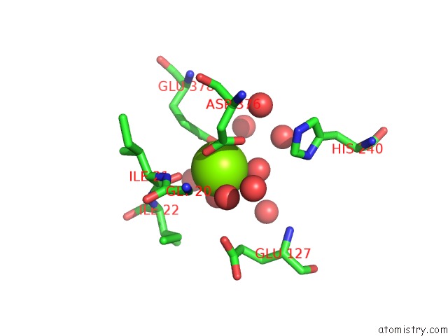
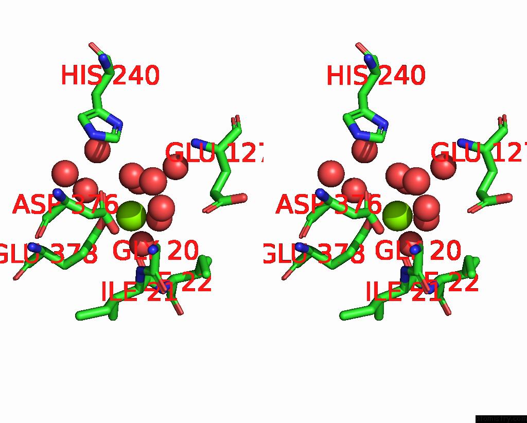
Magnesium binding site 1 out of 3 in 6mgl
Go back to
Magnesium binding site 1 out
of 3 in the Crystal Structure of the Catalytic Domain From GH74 Enzyme POGH74 From Paenibacillus Odorifer, D60A Mutant in Complex with Xxlg and Xgxxlg Xyloglucan
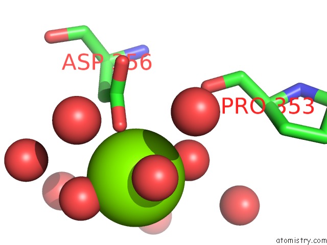
Mono view
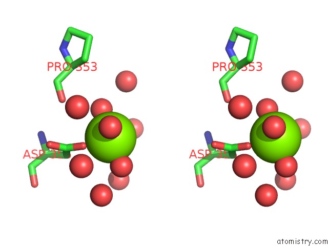
Stereo pair view
Mono view
Stereo pair view
A full contact list of Magnesium with other atoms in the Mg binding
site number 1 of Crystal Structure of the Catalytic Domain From GH74 Enzyme POGH74 From Paenibacillus Odorifer, D60A Mutant in Complex with Xxlg and Xgxxlg Xyloglucan within 5.0Å range:
|
Magnesium binding site 2 out of 3 in 6mgl
Go back to
Magnesium binding site 2 out
of 3 in the Crystal Structure of the Catalytic Domain From GH74 Enzyme POGH74 From Paenibacillus Odorifer, D60A Mutant in Complex with Xxlg and Xgxxlg Xyloglucan
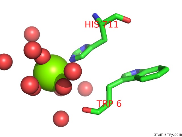
Mono view
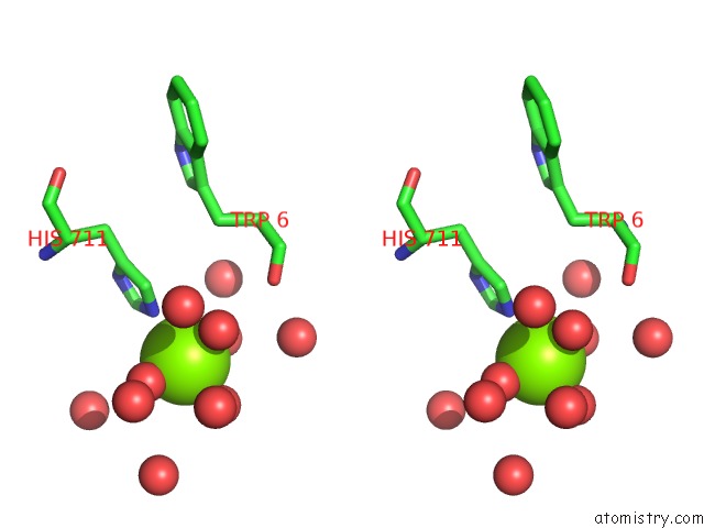
Stereo pair view
Mono view
Stereo pair view
A full contact list of Magnesium with other atoms in the Mg binding
site number 2 of Crystal Structure of the Catalytic Domain From GH74 Enzyme POGH74 From Paenibacillus Odorifer, D60A Mutant in Complex with Xxlg and Xgxxlg Xyloglucan within 5.0Å range:
|
Magnesium binding site 3 out of 3 in 6mgl
Go back to
Magnesium binding site 3 out
of 3 in the Crystal Structure of the Catalytic Domain From GH74 Enzyme POGH74 From Paenibacillus Odorifer, D60A Mutant in Complex with Xxlg and Xgxxlg Xyloglucan
Mono view
Stereo pair view
Mono view
Stereo pair view
A full contact list of Magnesium with other atoms in the Mg binding
site number 3 of Crystal Structure of the Catalytic Domain From GH74 Enzyme POGH74 From Paenibacillus Odorifer, D60A Mutant in Complex with Xxlg and Xgxxlg Xyloglucan within 5.0Å range:
|
Reference:
G.Arnal,
P.J.Stogios,
J.Asohan,
T.Skarina,
A.Savchenko,
H.Brumer.
Structural Enzymology Reveals the Molecular Basis of Substrate Regiospecificity and Processivity of An Exemplar Bacterial Glycoside Hydrolase Family 74ENDO-Xyloglucanase. Biochem. J. V. 475 3963 2018.
ISSN: ESSN 1470-8728
PubMed: 30463871
DOI: 10.1042/BCJ20180763
Page generated: Tue Oct 1 11:39:08 2024
ISSN: ESSN 1470-8728
PubMed: 30463871
DOI: 10.1042/BCJ20180763
Last articles
Zn in 9JYWZn in 9IR4
Zn in 9IR3
Zn in 9GMX
Zn in 9GMW
Zn in 9JEJ
Zn in 9ERF
Zn in 9ERE
Zn in 9EGV
Zn in 9EGW