Magnesium »
PDB 6m65-6mlq »
6mlh »
Magnesium in PDB 6mlh: Crystal Structure of X. Citri Phosphoglucomutase in Complex with Glucopyranosyl-1-Methyl-Phosphonic Acid
Protein crystallography data
The structure of Crystal Structure of X. Citri Phosphoglucomutase in Complex with Glucopyranosyl-1-Methyl-Phosphonic Acid, PDB code: 6mlh
was solved by
L.Beamer,
K.Stiers,
with X-Ray Crystallography technique. A brief refinement statistics is given in the table below:
| Resolution Low / High (Å) | 46.22 / 1.65 |
| Space group | P 21 21 21 |
| Cell size a, b, c (Å), α, β, γ (°) | 43.571, 54.715, 172.682, 90.00, 90.00, 90.00 |
| R / Rfree (%) | 17.2 / 21.3 |
Magnesium Binding Sites:
The binding sites of Magnesium atom in the Crystal Structure of X. Citri Phosphoglucomutase in Complex with Glucopyranosyl-1-Methyl-Phosphonic Acid
(pdb code 6mlh). This binding sites where shown within
5.0 Angstroms radius around Magnesium atom.
In total only one binding site of Magnesium was determined in the Crystal Structure of X. Citri Phosphoglucomutase in Complex with Glucopyranosyl-1-Methyl-Phosphonic Acid, PDB code: 6mlh:
In total only one binding site of Magnesium was determined in the Crystal Structure of X. Citri Phosphoglucomutase in Complex with Glucopyranosyl-1-Methyl-Phosphonic Acid, PDB code: 6mlh:
Magnesium binding site 1 out of 1 in 6mlh
Go back to
Magnesium binding site 1 out
of 1 in the Crystal Structure of X. Citri Phosphoglucomutase in Complex with Glucopyranosyl-1-Methyl-Phosphonic Acid
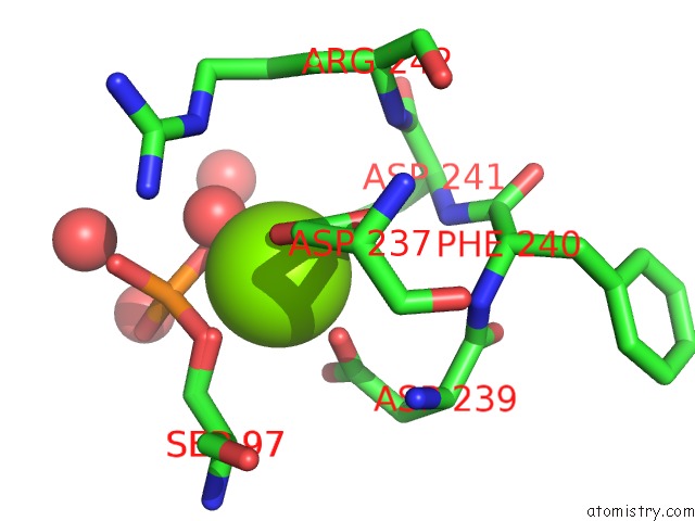
Mono view
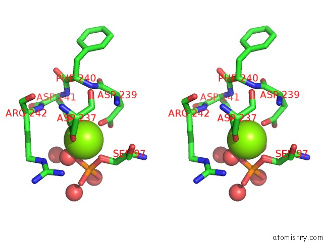
Stereo pair view
Mono view
Stereo pair view
A full contact list of Magnesium with other atoms in the Mg binding
site number 1 of Crystal Structure of X. Citri Phosphoglucomutase in Complex with Glucopyranosyl-1-Methyl-Phosphonic Acid within 5.0Å range:
|
Reference:
J.S.Zhu,
K.M.Stiers,
E.Soleimani,
B.R.Groves,
L.J.Beamer,
D.L.Jakeman.
Inhibitory Evaluation of Alpha Pmm/Pgm Frompseudomonas Aeruginosa: Chemical Synthesis, Enzyme Kinetics, and Protein Crystallographic Study. J.Org.Chem. V. 84 9627 2019.
ISSN: ISSN 0022-3263
PubMed: 31264865
DOI: 10.1021/ACS.JOC.9B01305
Page generated: Tue Oct 1 11:41:38 2024
ISSN: ISSN 0022-3263
PubMed: 31264865
DOI: 10.1021/ACS.JOC.9B01305
Last articles
Zn in 9J0NZn in 9J0O
Zn in 9J0P
Zn in 9FJX
Zn in 9EKB
Zn in 9C0F
Zn in 9CAH
Zn in 9CH0
Zn in 9CH3
Zn in 9CH1