Magnesium »
PDB 6mxg-6n7e »
6mxg »
Magnesium in PDB 6mxg: Crystal Structure of Trypanosoma Brucei Hypoxanthine-Guanine Phosphoribosyltranferase in Complex with Xmp
Enzymatic activity of Crystal Structure of Trypanosoma Brucei Hypoxanthine-Guanine Phosphoribosyltranferase in Complex with Xmp
All present enzymatic activity of Crystal Structure of Trypanosoma Brucei Hypoxanthine-Guanine Phosphoribosyltranferase in Complex with Xmp:
2.4.2.8;
2.4.2.8;
Protein crystallography data
The structure of Crystal Structure of Trypanosoma Brucei Hypoxanthine-Guanine Phosphoribosyltranferase in Complex with Xmp, PDB code: 6mxg
was solved by
D.Teran,
L.Guddat,
with X-Ray Crystallography technique. A brief refinement statistics is given in the table below:
| Resolution Low / High (Å) | 47.19 / 2.39 |
| Space group | C 1 2 1 |
| Cell size a, b, c (Å), α, β, γ (°) | 117.570, 94.370, 45.000, 90.00, 111.97, 90.00 |
| R / Rfree (%) | 25.7 / 28.5 |
Magnesium Binding Sites:
The binding sites of Magnesium atom in the Crystal Structure of Trypanosoma Brucei Hypoxanthine-Guanine Phosphoribosyltranferase in Complex with Xmp
(pdb code 6mxg). This binding sites where shown within
5.0 Angstroms radius around Magnesium atom.
In total 4 binding sites of Magnesium where determined in the Crystal Structure of Trypanosoma Brucei Hypoxanthine-Guanine Phosphoribosyltranferase in Complex with Xmp, PDB code: 6mxg:
Jump to Magnesium binding site number: 1; 2; 3; 4;
In total 4 binding sites of Magnesium where determined in the Crystal Structure of Trypanosoma Brucei Hypoxanthine-Guanine Phosphoribosyltranferase in Complex with Xmp, PDB code: 6mxg:
Jump to Magnesium binding site number: 1; 2; 3; 4;
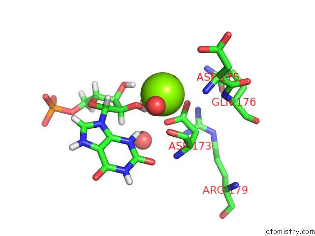
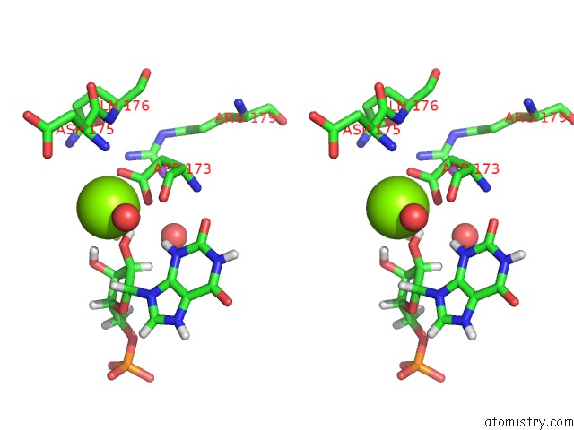
Magnesium binding site 1 out of 4 in 6mxg
Go back to
Magnesium binding site 1 out
of 4 in the Crystal Structure of Trypanosoma Brucei Hypoxanthine-Guanine Phosphoribosyltranferase in Complex with Xmp
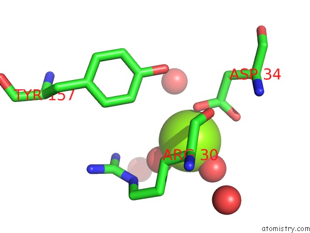
Mono view
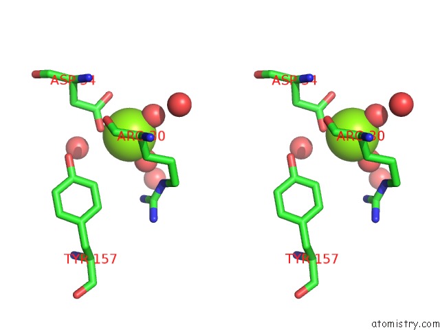
Stereo pair view
Mono view
Stereo pair view
A full contact list of Magnesium with other atoms in the Mg binding
site number 1 of Crystal Structure of Trypanosoma Brucei Hypoxanthine-Guanine Phosphoribosyltranferase in Complex with Xmp within 5.0Å range:
|
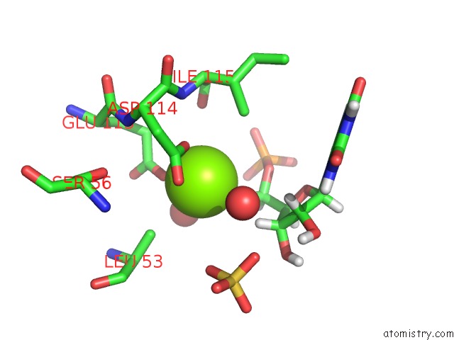
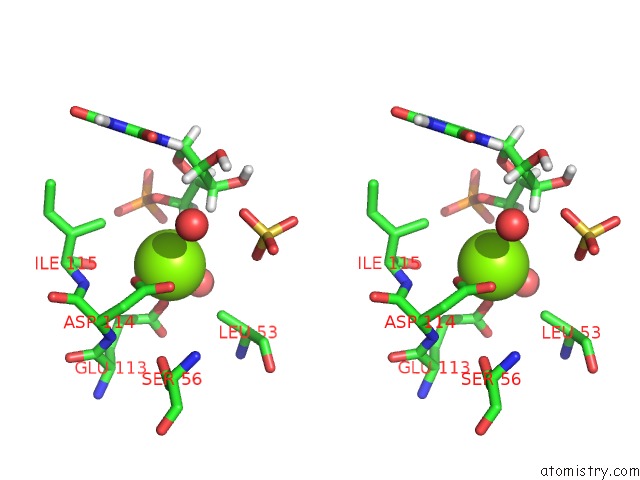
Magnesium binding site 2 out of 4 in 6mxg
Go back to
Magnesium binding site 2 out
of 4 in the Crystal Structure of Trypanosoma Brucei Hypoxanthine-Guanine Phosphoribosyltranferase in Complex with Xmp
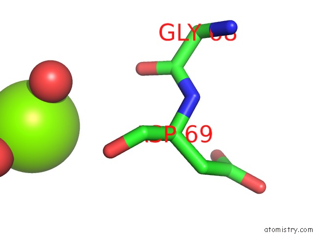
Mono view
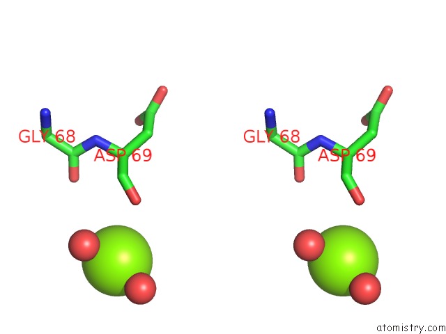
Stereo pair view
Mono view
Stereo pair view
A full contact list of Magnesium with other atoms in the Mg binding
site number 2 of Crystal Structure of Trypanosoma Brucei Hypoxanthine-Guanine Phosphoribosyltranferase in Complex with Xmp within 5.0Å range:
|
Magnesium binding site 3 out of 4 in 6mxg
Go back to
Magnesium binding site 3 out
of 4 in the Crystal Structure of Trypanosoma Brucei Hypoxanthine-Guanine Phosphoribosyltranferase in Complex with Xmp
Mono view
Stereo pair view
Mono view
Stereo pair view
A full contact list of Magnesium with other atoms in the Mg binding
site number 3 of Crystal Structure of Trypanosoma Brucei Hypoxanthine-Guanine Phosphoribosyltranferase in Complex with Xmp within 5.0Å range:
|
Magnesium binding site 4 out of 4 in 6mxg
Go back to
Magnesium binding site 4 out
of 4 in the Crystal Structure of Trypanosoma Brucei Hypoxanthine-Guanine Phosphoribosyltranferase in Complex with Xmp
Mono view
Stereo pair view
Mono view
Stereo pair view
A full contact list of Magnesium with other atoms in the Mg binding
site number 4 of Crystal Structure of Trypanosoma Brucei Hypoxanthine-Guanine Phosphoribosyltranferase in Complex with Xmp within 5.0Å range:
|
Reference:
D.Teran,
E.Dolezelova,
D.T.Keough,
D.Hockova,
A.Zikova,
L.W.Guddat.
Crystal Structures of Trypanosoma Brucei Hypoxanthine - Guanine - Xanthine Phosphoribosyltransferase in Complex with Imp, Gmp and Xmp. Febs J. 2019.
ISSN: ISSN 1742-464X
PubMed: 31287615
DOI: 10.1111/FEBS.14987
Page generated: Tue Oct 1 12:22:14 2024
ISSN: ISSN 1742-464X
PubMed: 31287615
DOI: 10.1111/FEBS.14987
Last articles
Zn in 9J0NZn in 9J0O
Zn in 9J0P
Zn in 9FJX
Zn in 9EKB
Zn in 9C0F
Zn in 9CAH
Zn in 9CH0
Zn in 9CH3
Zn in 9CH1