Magnesium »
PDB 6mxo-6n7i »
6n2t »
Magnesium in PDB 6n2t: Ternary Complex Crystal Structure of Dna Polymerase Beta with 5- Hydroxymethyl-Dc (5-Hmc) at the Templating Position
Enzymatic activity of Ternary Complex Crystal Structure of Dna Polymerase Beta with 5- Hydroxymethyl-Dc (5-Hmc) at the Templating Position
All present enzymatic activity of Ternary Complex Crystal Structure of Dna Polymerase Beta with 5- Hydroxymethyl-Dc (5-Hmc) at the Templating Position:
2.7.7.7;
2.7.7.7;
Protein crystallography data
The structure of Ternary Complex Crystal Structure of Dna Polymerase Beta with 5- Hydroxymethyl-Dc (5-Hmc) at the Templating Position, PDB code: 6n2t
was solved by
V.K.Batra,
S.H.Wilson,
with X-Ray Crystallography technique. A brief refinement statistics is given in the table below:
| Resolution Low / High (Å) | 28.90 / 2.60 |
| Space group | P 1 21 1 |
| Cell size a, b, c (Å), α, β, γ (°) | 50.476, 79.089, 55.556, 90.00, 106.61, 90.00 |
| R / Rfree (%) | 19.8 / 29.1 |
Other elements in 6n2t:
The structure of Ternary Complex Crystal Structure of Dna Polymerase Beta with 5- Hydroxymethyl-Dc (5-Hmc) at the Templating Position also contains other interesting chemical elements:
| Sodium | (Na) | 2 atoms |
Magnesium Binding Sites:
The binding sites of Magnesium atom in the Ternary Complex Crystal Structure of Dna Polymerase Beta with 5- Hydroxymethyl-Dc (5-Hmc) at the Templating Position
(pdb code 6n2t). This binding sites where shown within
5.0 Angstroms radius around Magnesium atom.
In total 2 binding sites of Magnesium where determined in the Ternary Complex Crystal Structure of Dna Polymerase Beta with 5- Hydroxymethyl-Dc (5-Hmc) at the Templating Position, PDB code: 6n2t:
Jump to Magnesium binding site number: 1; 2;
In total 2 binding sites of Magnesium where determined in the Ternary Complex Crystal Structure of Dna Polymerase Beta with 5- Hydroxymethyl-Dc (5-Hmc) at the Templating Position, PDB code: 6n2t:
Jump to Magnesium binding site number: 1; 2;
Magnesium binding site 1 out of 2 in 6n2t
Go back to
Magnesium binding site 1 out
of 2 in the Ternary Complex Crystal Structure of Dna Polymerase Beta with 5- Hydroxymethyl-Dc (5-Hmc) at the Templating Position
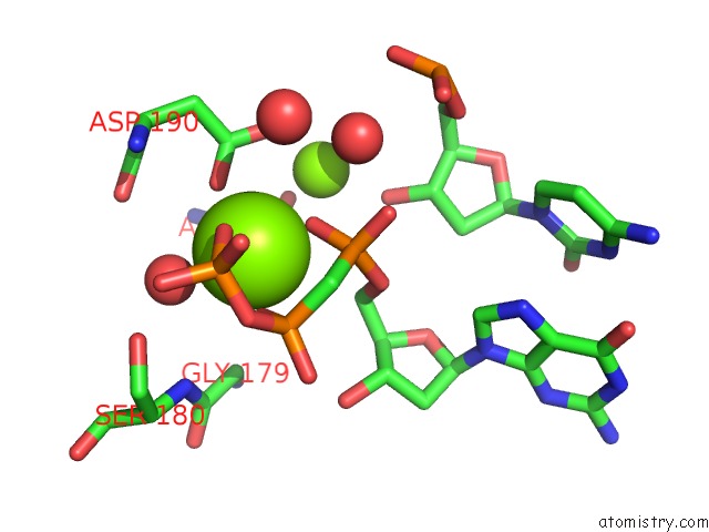
Mono view
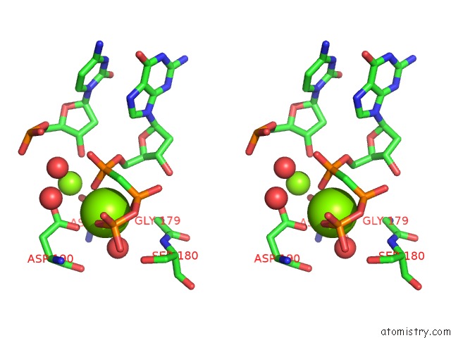
Stereo pair view
Mono view
Stereo pair view
A full contact list of Magnesium with other atoms in the Mg binding
site number 1 of Ternary Complex Crystal Structure of Dna Polymerase Beta with 5- Hydroxymethyl-Dc (5-Hmc) at the Templating Position within 5.0Å range:
|
Magnesium binding site 2 out of 2 in 6n2t
Go back to
Magnesium binding site 2 out
of 2 in the Ternary Complex Crystal Structure of Dna Polymerase Beta with 5- Hydroxymethyl-Dc (5-Hmc) at the Templating Position
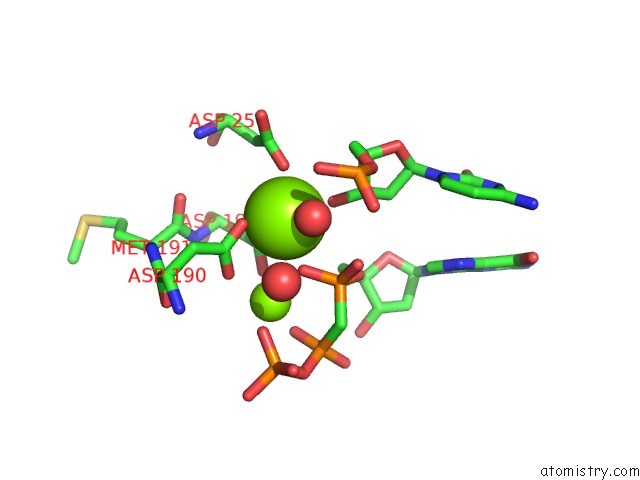
Mono view
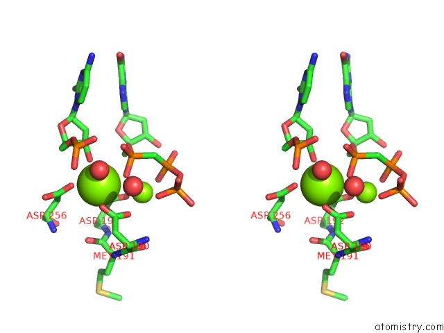
Stereo pair view
Mono view
Stereo pair view
A full contact list of Magnesium with other atoms in the Mg binding
site number 2 of Ternary Complex Crystal Structure of Dna Polymerase Beta with 5- Hydroxymethyl-Dc (5-Hmc) at the Templating Position within 5.0Å range:
|
Reference:
M.J.Howard,
K.G.Foley,
D.D.Shock,
V.K.Batra,
S.H.Wilson.
Molecular Basis For the Faithful Replication of 5-Methylcytosine and Its Oxidized Forms By Dna Polymerase Beta. J.Biol.Chem. V. 294 7194 2019.
ISSN: ESSN 1083-351X
PubMed: 30885943
DOI: 10.1074/JBC.RA118.006809
Page generated: Tue Oct 1 12:24:32 2024
ISSN: ESSN 1083-351X
PubMed: 30885943
DOI: 10.1074/JBC.RA118.006809
Last articles
Zn in 9MJ5Zn in 9HNW
Zn in 9G0L
Zn in 9FNE
Zn in 9DZN
Zn in 9E0I
Zn in 9D32
Zn in 9DAK
Zn in 8ZXC
Zn in 8ZUF