Magnesium »
PDB 6mxo-6n7i »
6n62 »
Magnesium in PDB 6n62: Escherichia Coli Rna Polymerase SIGMA70-Holoenzyme Bound to Upstream Fork Promoter Dna
Enzymatic activity of Escherichia Coli Rna Polymerase SIGMA70-Holoenzyme Bound to Upstream Fork Promoter Dna
All present enzymatic activity of Escherichia Coli Rna Polymerase SIGMA70-Holoenzyme Bound to Upstream Fork Promoter Dna:
2.7.7.6;
2.7.7.6;
Protein crystallography data
The structure of Escherichia Coli Rna Polymerase SIGMA70-Holoenzyme Bound to Upstream Fork Promoter Dna, PDB code: 6n62
was solved by
N.Braffman,
J.Hauver,
E.A.Campbell,
S.A.Darst,
with X-Ray Crystallography technique. A brief refinement statistics is given in the table below:
| Resolution Low / High (Å) | 24.88 / 3.80 |
| Space group | P 41 21 2 |
| Cell size a, b, c (Å), α, β, γ (°) | 173.771, 173.771, 388.771, 90.00, 90.00, 90.00 |
| R / Rfree (%) | 28.6 / 33.4 |
Other elements in 6n62:
The structure of Escherichia Coli Rna Polymerase SIGMA70-Holoenzyme Bound to Upstream Fork Promoter Dna also contains other interesting chemical elements:
| Zinc | (Zn) | 2 atoms |
Magnesium Binding Sites:
The binding sites of Magnesium atom in the Escherichia Coli Rna Polymerase SIGMA70-Holoenzyme Bound to Upstream Fork Promoter Dna
(pdb code 6n62). This binding sites where shown within
5.0 Angstroms radius around Magnesium atom.
In total only one binding site of Magnesium was determined in the Escherichia Coli Rna Polymerase SIGMA70-Holoenzyme Bound to Upstream Fork Promoter Dna, PDB code: 6n62:
In total only one binding site of Magnesium was determined in the Escherichia Coli Rna Polymerase SIGMA70-Holoenzyme Bound to Upstream Fork Promoter Dna, PDB code: 6n62:
Magnesium binding site 1 out of 1 in 6n62
Go back to
Magnesium binding site 1 out
of 1 in the Escherichia Coli Rna Polymerase SIGMA70-Holoenzyme Bound to Upstream Fork Promoter Dna
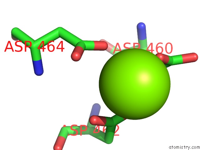
Mono view
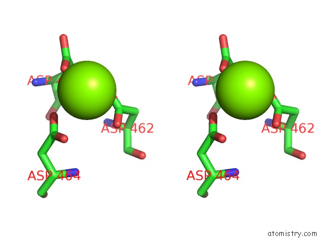
Stereo pair view
Mono view
Stereo pair view
A full contact list of Magnesium with other atoms in the Mg binding
site number 1 of Escherichia Coli Rna Polymerase SIGMA70-Holoenzyme Bound to Upstream Fork Promoter Dna within 5.0Å range:
|
Reference:
N.R.Braffman,
F.J.Piscotta,
J.Hauver,
E.A.Campbell,
A.J.Link,
S.A.Darst.
Structural Mechanism of Transcription Inhibition By Lasso Peptides Microcin J25 and Capistruin. Proc. Natl. Acad. Sci. V. 116 1273 2019U.S.A..
ISSN: ESSN 1091-6490
PubMed: 30626643
DOI: 10.1073/PNAS.1817352116
Page generated: Tue Oct 1 12:30:38 2024
ISSN: ESSN 1091-6490
PubMed: 30626643
DOI: 10.1073/PNAS.1817352116
Last articles
Zn in 9MJ5Zn in 9HNW
Zn in 9G0L
Zn in 9FNE
Zn in 9DZN
Zn in 9E0I
Zn in 9D32
Zn in 9DAK
Zn in 8ZXC
Zn in 8ZUF