Magnesium »
PDB 6n7n-6nk8 »
6njd »
Magnesium in PDB 6njd: Crystal Structure of Ravd (Residues 1-200) From Legionella Pneumophila (Strain Corby) Complexed with Met-1 Linked Di-Ubiquitin
Protein crystallography data
The structure of Crystal Structure of Ravd (Residues 1-200) From Legionella Pneumophila (Strain Corby) Complexed with Met-1 Linked Di-Ubiquitin, PDB code: 6njd
was solved by
X.Wang,
Y.Zhou,
Y.Zhu,
with X-Ray Crystallography technique. A brief refinement statistics is given in the table below:
| Resolution Low / High (Å) | 50.01 / 2.05 |
| Space group | P 21 21 2 |
| Cell size a, b, c (Å), α, β, γ (°) | 77.740, 102.090, 90.920, 90.00, 90.00, 90.00 |
| R / Rfree (%) | 21.2 / 24.6 |
Magnesium Binding Sites:
The binding sites of Magnesium atom in the Crystal Structure of Ravd (Residues 1-200) From Legionella Pneumophila (Strain Corby) Complexed with Met-1 Linked Di-Ubiquitin
(pdb code 6njd). This binding sites where shown within
5.0 Angstroms radius around Magnesium atom.
In total 2 binding sites of Magnesium where determined in the Crystal Structure of Ravd (Residues 1-200) From Legionella Pneumophila (Strain Corby) Complexed with Met-1 Linked Di-Ubiquitin, PDB code: 6njd:
Jump to Magnesium binding site number: 1; 2;
In total 2 binding sites of Magnesium where determined in the Crystal Structure of Ravd (Residues 1-200) From Legionella Pneumophila (Strain Corby) Complexed with Met-1 Linked Di-Ubiquitin, PDB code: 6njd:
Jump to Magnesium binding site number: 1; 2;
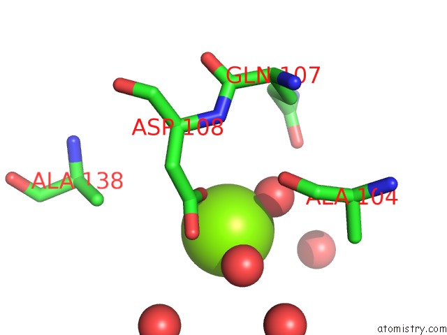
Magnesium binding site 1 out of 2 in 6njd
Go back to
Magnesium binding site 1 out
of 2 in the Crystal Structure of Ravd (Residues 1-200) From Legionella Pneumophila (Strain Corby) Complexed with Met-1 Linked Di-Ubiquitin
Mono view

Stereo pair view
Mono view
Stereo pair view
A full contact list of Magnesium with other atoms in the Mg binding
site number 1 of Crystal Structure of Ravd (Residues 1-200) From Legionella Pneumophila (Strain Corby) Complexed with Met-1 Linked Di-Ubiquitin within 5.0Å range:
|
Magnesium binding site 2 out of 2 in 6njd
Go back to
Magnesium binding site 2 out
of 2 in the Crystal Structure of Ravd (Residues 1-200) From Legionella Pneumophila (Strain Corby) Complexed with Met-1 Linked Di-Ubiquitin

Mono view

Stereo pair view
Mono view
Stereo pair view
A full contact list of Magnesium with other atoms in the Mg binding
site number 2 of Crystal Structure of Ravd (Residues 1-200) From Legionella Pneumophila (Strain Corby) Complexed with Met-1 Linked Di-Ubiquitin within 5.0Å range:
|
Reference:
M.Wan,
X.Wang,
C.Huang,
D.Xu,
Z.Wang,
Y.Zhou,
Y.Zhu.
A Bacterial Effector Deubiquitinase Specifically Hydrolyses Linear Ubiquitin Chains to Inhibit Host Inflammatory Signalling. Nat Microbiol V. 4 1282 2019.
ISSN: ESSN 2058-5276
PubMed: 31110362
DOI: 10.1038/S41564-019-0454-1
Page generated: Tue Oct 1 12:40:24 2024
ISSN: ESSN 2058-5276
PubMed: 31110362
DOI: 10.1038/S41564-019-0454-1
Last articles
Zn in 9MJ5Zn in 9HNW
Zn in 9G0L
Zn in 9FNE
Zn in 9DZN
Zn in 9E0I
Zn in 9D32
Zn in 9DAK
Zn in 8ZXC
Zn in 8ZUF