Magnesium »
PDB 6n7i-6njw »
6njj »
Magnesium in PDB 6njj: Crystal Structure of the PDE4D Catalytic Domain and UCR2 Regulatory Helix with BPN14770
Enzymatic activity of Crystal Structure of the PDE4D Catalytic Domain and UCR2 Regulatory Helix with BPN14770
All present enzymatic activity of Crystal Structure of the PDE4D Catalytic Domain and UCR2 Regulatory Helix with BPN14770:
3.1.4.53;
3.1.4.53;
Protein crystallography data
The structure of Crystal Structure of the PDE4D Catalytic Domain and UCR2 Regulatory Helix with BPN14770, PDB code: 6njj
was solved by
D.Fox Iii,
J.W.Fairman,
M.E.Gurney,
with X-Ray Crystallography technique. A brief refinement statistics is given in the table below:
| Resolution Low / High (Å) | 45.49 / 2.30 |
| Space group | P 1 21 1 |
| Cell size a, b, c (Å), α, β, γ (°) | 81.330, 82.050, 116.350, 90.00, 110.01, 90.00 |
| R / Rfree (%) | 16.7 / 21.4 |
Other elements in 6njj:
The structure of Crystal Structure of the PDE4D Catalytic Domain and UCR2 Regulatory Helix with BPN14770 also contains other interesting chemical elements:
| Fluorine | (F) | 12 atoms |
| Zinc | (Zn) | 6 atoms |
| Chlorine | (Cl) | 4 atoms |
Magnesium Binding Sites:
The binding sites of Magnesium atom in the Crystal Structure of the PDE4D Catalytic Domain and UCR2 Regulatory Helix with BPN14770
(pdb code 6njj). This binding sites where shown within
5.0 Angstroms radius around Magnesium atom.
In total 4 binding sites of Magnesium where determined in the Crystal Structure of the PDE4D Catalytic Domain and UCR2 Regulatory Helix with BPN14770, PDB code: 6njj:
Jump to Magnesium binding site number: 1; 2; 3; 4;
In total 4 binding sites of Magnesium where determined in the Crystal Structure of the PDE4D Catalytic Domain and UCR2 Regulatory Helix with BPN14770, PDB code: 6njj:
Jump to Magnesium binding site number: 1; 2; 3; 4;
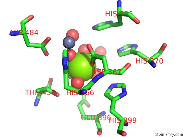
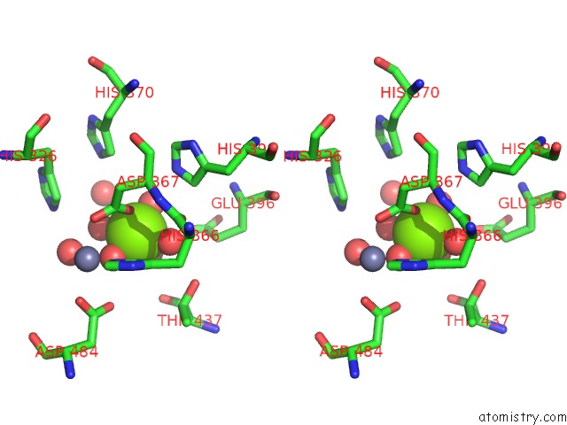
Magnesium binding site 1 out of 4 in 6njj
Go back to
Magnesium binding site 1 out
of 4 in the Crystal Structure of the PDE4D Catalytic Domain and UCR2 Regulatory Helix with BPN14770
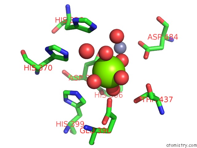
Mono view
Stereo pair view
Mono view
Stereo pair view
A full contact list of Magnesium with other atoms in the Mg binding
site number 1 of Crystal Structure of the PDE4D Catalytic Domain and UCR2 Regulatory Helix with BPN14770 within 5.0Å range:
|
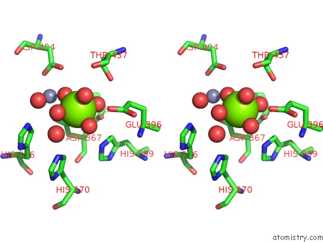
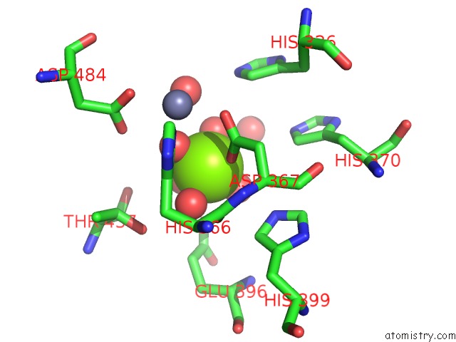
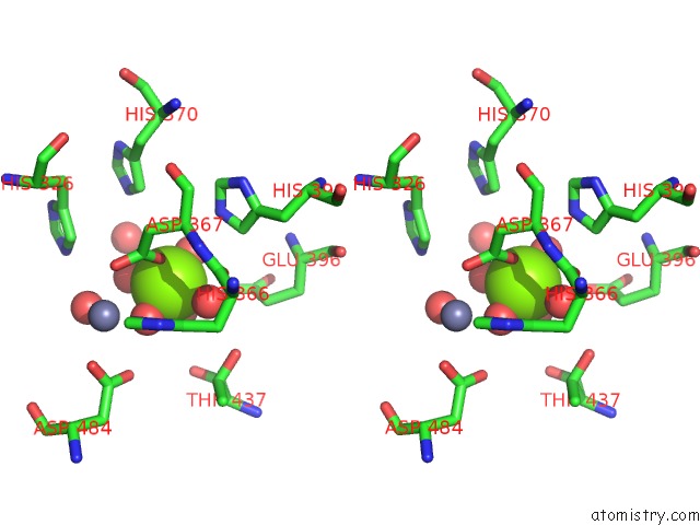
Magnesium binding site 2 out of 4 in 6njj
Go back to
Magnesium binding site 2 out
of 4 in the Crystal Structure of the PDE4D Catalytic Domain and UCR2 Regulatory Helix with BPN14770
Mono view
Stereo pair view
Mono view
Stereo pair view
A full contact list of Magnesium with other atoms in the Mg binding
site number 2 of Crystal Structure of the PDE4D Catalytic Domain and UCR2 Regulatory Helix with BPN14770 within 5.0Å range:
|
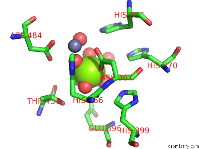
Magnesium binding site 3 out of 4 in 6njj
Go back to
Magnesium binding site 3 out
of 4 in the Crystal Structure of the PDE4D Catalytic Domain and UCR2 Regulatory Helix with BPN14770
Mono view
Stereo pair view
Mono view
Stereo pair view
A full contact list of Magnesium with other atoms in the Mg binding
site number 3 of Crystal Structure of the PDE4D Catalytic Domain and UCR2 Regulatory Helix with BPN14770 within 5.0Å range:
|
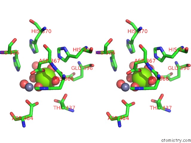
Magnesium binding site 4 out of 4 in 6njj
Go back to
Magnesium binding site 4 out
of 4 in the Crystal Structure of the PDE4D Catalytic Domain and UCR2 Regulatory Helix with BPN14770
Mono view
Stereo pair view
Mono view
Stereo pair view
A full contact list of Magnesium with other atoms in the Mg binding
site number 4 of Crystal Structure of the PDE4D Catalytic Domain and UCR2 Regulatory Helix with BPN14770 within 5.0Å range:
|
Reference:
M.E.Gurney,
R.A.Nugent,
X.Mo,
J.A.Sindac,
T.J.Hagen,
D.Fox 3Rd,
J.M.O'donnell,
C.Zhang,
Y.Xu,
H.T.Zhang,
V.E.Groppi,
M.Bailie,
R.E.White,
D.L.Romero,
A.S.Vellekoop,
J.R.Walker,
M.D.Surman,
L.Zhu,
R.F.Campbell.
Design and Synthesis of Selective Phosphodiesterase 4D (PDE4D) Allosteric Inhibitors For the Treatment of Fragile X Syndrome and Other Brain Disorders. J.Med.Chem. V. 62 4884 2019.
ISSN: ISSN 0022-2623
PubMed: 31013090
DOI: 10.1021/ACS.JMEDCHEM.9B00193
Page generated: Tue Oct 1 12:41:17 2024
ISSN: ISSN 0022-2623
PubMed: 31013090
DOI: 10.1021/ACS.JMEDCHEM.9B00193
Last articles
Zn in 9J0NZn in 9J0O
Zn in 9J0P
Zn in 9FJX
Zn in 9EKB
Zn in 9C0F
Zn in 9CAH
Zn in 9CH0
Zn in 9CH3
Zn in 9CH1