Magnesium »
PDB 6nop-6o2r »
6nop »
Magnesium in PDB 6nop: Structure of Cyanothece Mcda(D38A)-Atp Complex
Protein crystallography data
The structure of Structure of Cyanothece Mcda(D38A)-Atp Complex, PDB code: 6nop
was solved by
M.A.Schumacher,
with X-Ray Crystallography technique. A brief refinement statistics is given in the table below:
| Resolution Low / High (Å) | 60.87 / 1.70 |
| Space group | P 31 2 1 |
| Cell size a, b, c (Å), α, β, γ (°) | 115.658, 115.658, 76.648, 90.00, 90.00, 120.00 |
| R / Rfree (%) | 18.2 / 20.4 |
Magnesium Binding Sites:
The binding sites of Magnesium atom in the Structure of Cyanothece Mcda(D38A)-Atp Complex
(pdb code 6nop). This binding sites where shown within
5.0 Angstroms radius around Magnesium atom.
In total 2 binding sites of Magnesium where determined in the Structure of Cyanothece Mcda(D38A)-Atp Complex, PDB code: 6nop:
Jump to Magnesium binding site number: 1; 2;
In total 2 binding sites of Magnesium where determined in the Structure of Cyanothece Mcda(D38A)-Atp Complex, PDB code: 6nop:
Jump to Magnesium binding site number: 1; 2;
Magnesium binding site 1 out of 2 in 6nop
Go back to
Magnesium binding site 1 out
of 2 in the Structure of Cyanothece Mcda(D38A)-Atp Complex
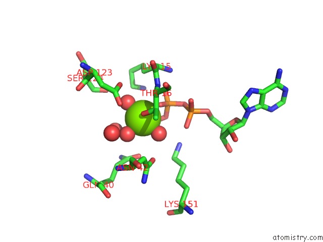
Mono view
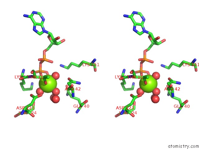
Stereo pair view
Mono view
Stereo pair view
A full contact list of Magnesium with other atoms in the Mg binding
site number 1 of Structure of Cyanothece Mcda(D38A)-Atp Complex within 5.0Å range:
|
Magnesium binding site 2 out of 2 in 6nop
Go back to
Magnesium binding site 2 out
of 2 in the Structure of Cyanothece Mcda(D38A)-Atp Complex
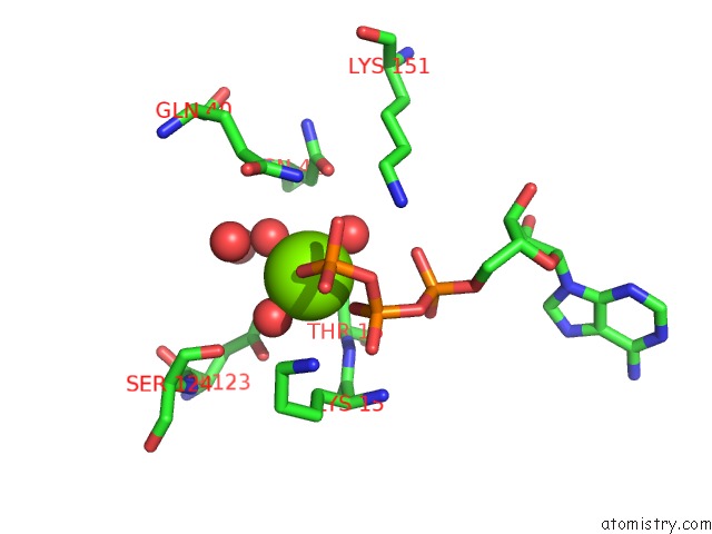
Mono view
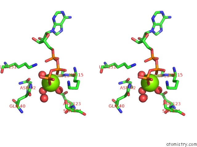
Stereo pair view
Mono view
Stereo pair view
A full contact list of Magnesium with other atoms in the Mg binding
site number 2 of Structure of Cyanothece Mcda(D38A)-Atp Complex within 5.0Å range:
|
Reference:
M.A.Schumacher,
M.Henderson,
H.Zhang.
Structures of Maintenance of Carboxysome Distribution Walker-Box Mcda and Mcdb Adaptor Homologs. Nucleic Acids Res. V. 47 5950 2019.
ISSN: ESSN 1362-4962
PubMed: 31106331
DOI: 10.1093/NAR/GKZ314
Page generated: Tue Oct 1 12:52:16 2024
ISSN: ESSN 1362-4962
PubMed: 31106331
DOI: 10.1093/NAR/GKZ314
Last articles
Ca in 5SVECa in 5SSX
Ca in 5SV0
Ca in 5STD
Ca in 5SSZ
Ca in 5SSY
Ca in 5SIC
Ca in 5SBD
Ca in 5SBE
Ca in 5SBC