Magnesium »
PDB 6nop-6o2r »
6np5 »
Magnesium in PDB 6np5: Aac-Via Bound to Kanamycin B
Enzymatic activity of Aac-Via Bound to Kanamycin B
All present enzymatic activity of Aac-Via Bound to Kanamycin B:
2.3.1.81;
2.3.1.81;
Protein crystallography data
The structure of Aac-Via Bound to Kanamycin B, PDB code: 6np5
was solved by
P.Kumar,
M.J.Cuneo,
with X-Ray Crystallography technique. A brief refinement statistics is given in the table below:
| Resolution Low / High (Å) | 43.21 / 1.35 |
| Space group | C 1 2 1 |
| Cell size a, b, c (Å), α, β, γ (°) | 86.746, 86.429, 50.119, 90.00, 119.75, 90.00 |
| R / Rfree (%) | 12.2 / 14 |
Magnesium Binding Sites:
The binding sites of Magnesium atom in the Aac-Via Bound to Kanamycin B
(pdb code 6np5). This binding sites where shown within
5.0 Angstroms radius around Magnesium atom.
In total 2 binding sites of Magnesium where determined in the Aac-Via Bound to Kanamycin B, PDB code: 6np5:
Jump to Magnesium binding site number: 1; 2;
In total 2 binding sites of Magnesium where determined in the Aac-Via Bound to Kanamycin B, PDB code: 6np5:
Jump to Magnesium binding site number: 1; 2;
Magnesium binding site 1 out of 2 in 6np5
Go back to
Magnesium binding site 1 out
of 2 in the Aac-Via Bound to Kanamycin B
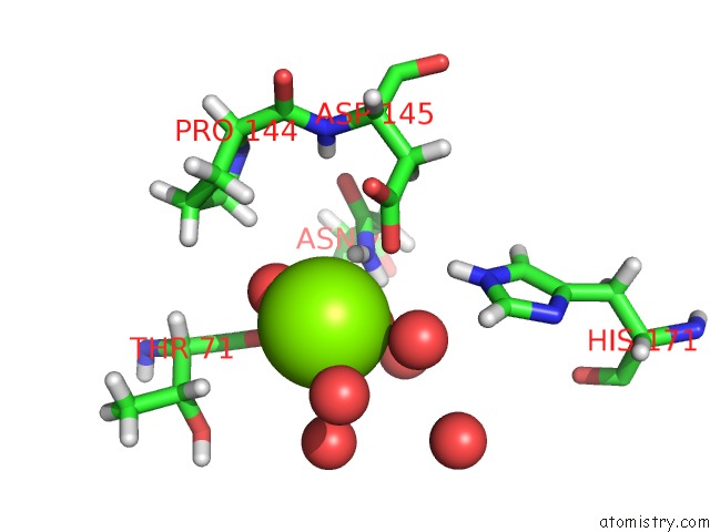
Mono view
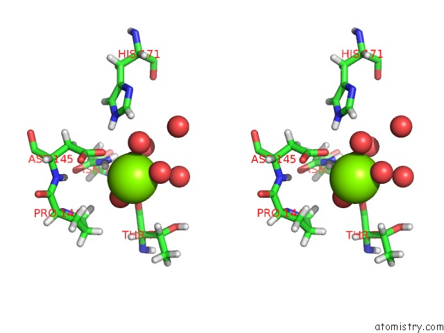
Stereo pair view
Mono view
Stereo pair view
A full contact list of Magnesium with other atoms in the Mg binding
site number 1 of Aac-Via Bound to Kanamycin B within 5.0Å range:
|
Magnesium binding site 2 out of 2 in 6np5
Go back to
Magnesium binding site 2 out
of 2 in the Aac-Via Bound to Kanamycin B
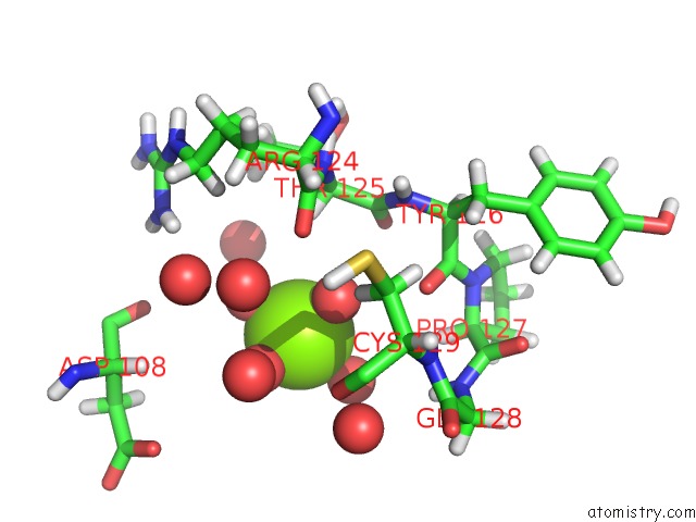
Mono view
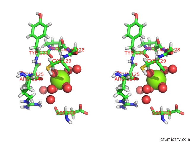
Stereo pair view
Mono view
Stereo pair view
A full contact list of Magnesium with other atoms in the Mg binding
site number 2 of Aac-Via Bound to Kanamycin B within 5.0Å range:
|
Reference:
P.Kumar,
P.K.Agarwal,
M.B.Waddell,
T.Mittag,
E.H.Serpersu,
M.J.Cuneo.
Low-Barrier and Canonical Hydrogen Bonds Modulate Activity and Specificity of A Catalytic Triad. Angew.Chem.Int.Ed.Engl. V. 58 16260 2019.
ISSN: ESSN 1521-3773
PubMed: 31515870
DOI: 10.1002/ANIE.201908535
Page generated: Tue Oct 1 12:52:42 2024
ISSN: ESSN 1521-3773
PubMed: 31515870
DOI: 10.1002/ANIE.201908535
Last articles
Fe in 2YXOFe in 2YRS
Fe in 2YXC
Fe in 2YNM
Fe in 2YVJ
Fe in 2YP1
Fe in 2YU2
Fe in 2YU1
Fe in 2YQB
Fe in 2YOO