Magnesium »
PDB 6nop-6o2r »
6nti »
Magnesium in PDB 6nti: Neutron/X-Ray Crystal Structure of Aac-Via Bound to Kanamycin B
Enzymatic activity of Neutron/X-Ray Crystal Structure of Aac-Via Bound to Kanamycin B
All present enzymatic activity of Neutron/X-Ray Crystal Structure of Aac-Via Bound to Kanamycin B:
2.3.1.81;
2.3.1.81;
Protein crystallography data
The structure of Neutron/X-Ray Crystal Structure of Aac-Via Bound to Kanamycin B, PDB code: 6nti
was solved by
M.J.Cuneo,
P.Kumar,
with X-Ray Crystallography technique. A brief refinement statistics is given in the table below:
| Resolution Low / High (Å) | N/A / 2.30 |
| Space group | I 1 2 1 |
| Cell size a, b, c (Å), α, β, γ (°) | 51.359, 86.058, 77.597, 90.00, 93.98, 90.00 |
| R / Rfree (%) | n/a / n/a |
Magnesium Binding Sites:
The binding sites of Magnesium atom in the Neutron/X-Ray Crystal Structure of Aac-Via Bound to Kanamycin B
(pdb code 6nti). This binding sites where shown within
5.0 Angstroms radius around Magnesium atom.
In total only one binding site of Magnesium was determined in the Neutron/X-Ray Crystal Structure of Aac-Via Bound to Kanamycin B, PDB code: 6nti:
In total only one binding site of Magnesium was determined in the Neutron/X-Ray Crystal Structure of Aac-Via Bound to Kanamycin B, PDB code: 6nti:
Magnesium binding site 1 out of 1 in 6nti
Go back to
Magnesium binding site 1 out
of 1 in the Neutron/X-Ray Crystal Structure of Aac-Via Bound to Kanamycin B
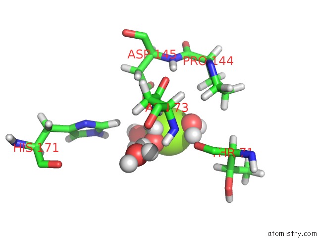
Mono view
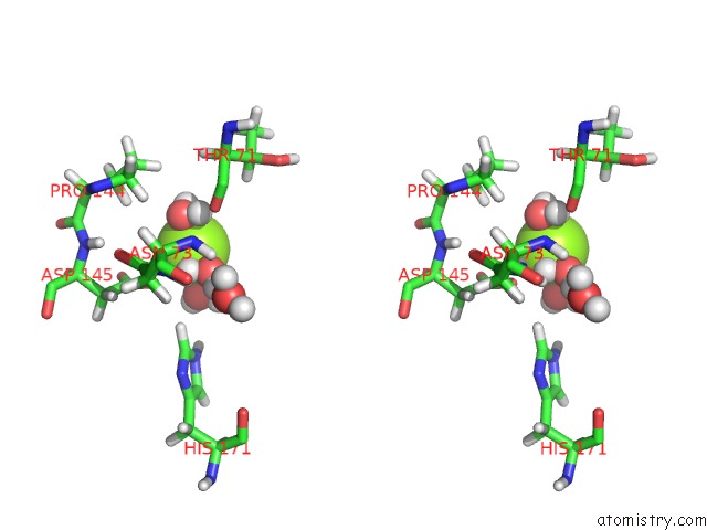
Stereo pair view
Mono view
Stereo pair view
A full contact list of Magnesium with other atoms in the Mg binding
site number 1 of Neutron/X-Ray Crystal Structure of Aac-Via Bound to Kanamycin B within 5.0Å range:
|
Reference:
P.Kumar,
P.K.Agarwal,
M.B.Waddell,
T.Mittag,
E.H.Serpersu,
M.J.Cuneo.
Low-Barrier and Canonical Hydrogen Bonds Modulate Activity and Specificity of A Catalytic Triad. Angew.Chem.Int.Ed.Engl. V. 58 16260 2019.
ISSN: ESSN 1521-3773
PubMed: 31515870
DOI: 10.1002/ANIE.201908535
Page generated: Tue Oct 1 12:53:42 2024
ISSN: ESSN 1521-3773
PubMed: 31515870
DOI: 10.1002/ANIE.201908535
Last articles
Zn in 9MJ5Zn in 9HNW
Zn in 9G0L
Zn in 9FNE
Zn in 9DZN
Zn in 9E0I
Zn in 9D32
Zn in 9DAK
Zn in 8ZXC
Zn in 8ZUF