Magnesium »
PDB 6o2r-6oiw »
6obj »
Magnesium in PDB 6obj: Structure of A Dna-Bound Dimer Extracted From Filamentous Sgrai Endonuclease in Its Activated Form
Magnesium Binding Sites:
The binding sites of Magnesium atom in the Structure of A Dna-Bound Dimer Extracted From Filamentous Sgrai Endonuclease in Its Activated Form
(pdb code 6obj). This binding sites where shown within
5.0 Angstroms radius around Magnesium atom.
In total 2 binding sites of Magnesium where determined in the Structure of A Dna-Bound Dimer Extracted From Filamentous Sgrai Endonuclease in Its Activated Form, PDB code: 6obj:
Jump to Magnesium binding site number: 1; 2;
In total 2 binding sites of Magnesium where determined in the Structure of A Dna-Bound Dimer Extracted From Filamentous Sgrai Endonuclease in Its Activated Form, PDB code: 6obj:
Jump to Magnesium binding site number: 1; 2;
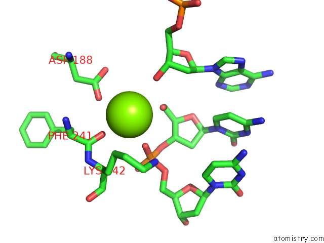
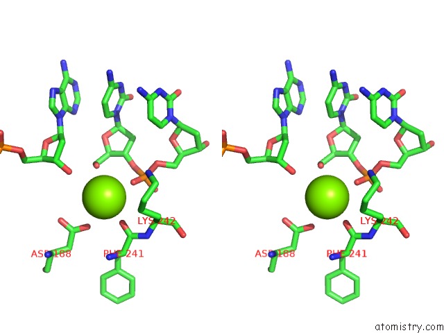
Magnesium binding site 1 out of 2 in 6obj
Go back to
Magnesium binding site 1 out
of 2 in the Structure of A Dna-Bound Dimer Extracted From Filamentous Sgrai Endonuclease in Its Activated Form

Mono view

Stereo pair view
Mono view
Stereo pair view
A full contact list of Magnesium with other atoms in the Mg binding
site number 1 of Structure of A Dna-Bound Dimer Extracted From Filamentous Sgrai Endonuclease in Its Activated Form within 5.0Å range:
|
Magnesium binding site 2 out of 2 in 6obj
Go back to
Magnesium binding site 2 out
of 2 in the Structure of A Dna-Bound Dimer Extracted From Filamentous Sgrai Endonuclease in Its Activated Form
Mono view
Stereo pair view
Mono view
Stereo pair view
A full contact list of Magnesium with other atoms in the Mg binding
site number 2 of Structure of A Dna-Bound Dimer Extracted From Filamentous Sgrai Endonuclease in Its Activated Form within 5.0Å range:
|
Reference:
S.Polley,
D.Lyumkis,
N.C.Horton.
Structure of A Dna-Bound Dimer Extracted From Filamentous Sgrai Endonuclease in Its Activated Form To Be Published.
Page generated: Tue Oct 1 13:21:35 2024
Last articles
Zn in 9J0NZn in 9J0O
Zn in 9J0P
Zn in 9FJX
Zn in 9EKB
Zn in 9C0F
Zn in 9CAH
Zn in 9CH0
Zn in 9CH3
Zn in 9CH1