Magnesium »
PDB 6oqz-6p1o »
6p0b »
Magnesium in PDB 6p0b: Human Dna Ligase 1 (E346A/E592A) Bound to An Adenylated, Dideoxy Terminated Dna Nick with 200 Mm MG2+
Enzymatic activity of Human Dna Ligase 1 (E346A/E592A) Bound to An Adenylated, Dideoxy Terminated Dna Nick with 200 Mm MG2+
All present enzymatic activity of Human Dna Ligase 1 (E346A/E592A) Bound to An Adenylated, Dideoxy Terminated Dna Nick with 200 Mm MG2+:
6.5.1.1;
6.5.1.1;
Protein crystallography data
The structure of Human Dna Ligase 1 (E346A/E592A) Bound to An Adenylated, Dideoxy Terminated Dna Nick with 200 Mm MG2+, PDB code: 6p0b
was solved by
M.J.Schellenberg,
R.S.Williams,
P.S.Tumbale,
A.A.Riccio,
with X-Ray Crystallography technique. A brief refinement statistics is given in the table below:
| Resolution Low / High (Å) | 41.43 / 2.20 |
| Space group | P 21 21 21 |
| Cell size a, b, c (Å), α, β, γ (°) | 71.866, 101.398, 115.606, 90.00, 90.00, 90.00 |
| R / Rfree (%) | 17 / 20.6 |
Magnesium Binding Sites:
The binding sites of Magnesium atom in the Human Dna Ligase 1 (E346A/E592A) Bound to An Adenylated, Dideoxy Terminated Dna Nick with 200 Mm MG2+
(pdb code 6p0b). This binding sites where shown within
5.0 Angstroms radius around Magnesium atom.
In total 4 binding sites of Magnesium where determined in the Human Dna Ligase 1 (E346A/E592A) Bound to An Adenylated, Dideoxy Terminated Dna Nick with 200 Mm MG2+, PDB code: 6p0b:
Jump to Magnesium binding site number: 1; 2; 3; 4;
In total 4 binding sites of Magnesium where determined in the Human Dna Ligase 1 (E346A/E592A) Bound to An Adenylated, Dideoxy Terminated Dna Nick with 200 Mm MG2+, PDB code: 6p0b:
Jump to Magnesium binding site number: 1; 2; 3; 4;
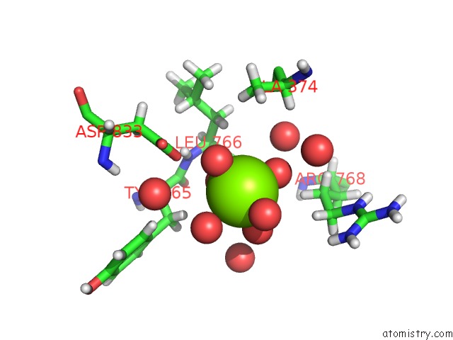
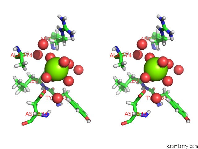
Magnesium binding site 1 out of 4 in 6p0b
Go back to
Magnesium binding site 1 out
of 4 in the Human Dna Ligase 1 (E346A/E592A) Bound to An Adenylated, Dideoxy Terminated Dna Nick with 200 Mm MG2+
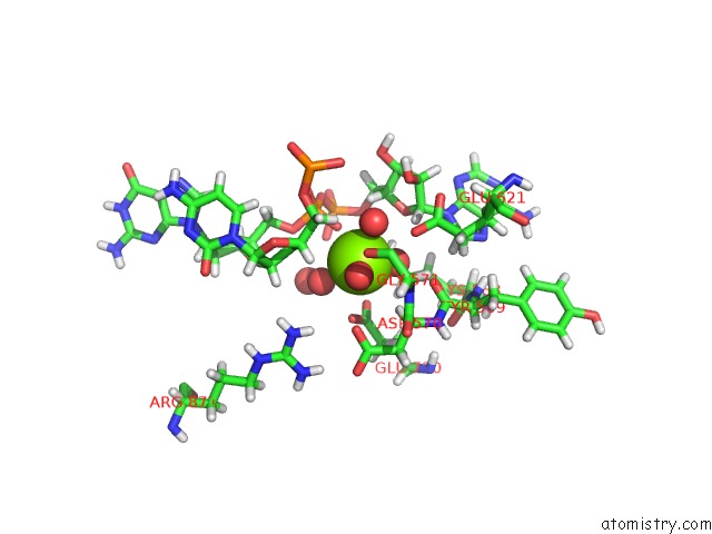
Mono view
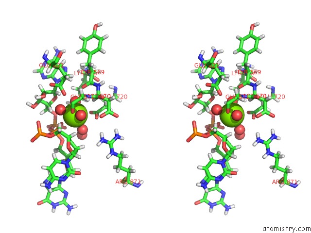
Stereo pair view
Mono view
Stereo pair view
A full contact list of Magnesium with other atoms in the Mg binding
site number 1 of Human Dna Ligase 1 (E346A/E592A) Bound to An Adenylated, Dideoxy Terminated Dna Nick with 200 Mm MG2+ within 5.0Å range:
|
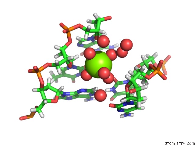
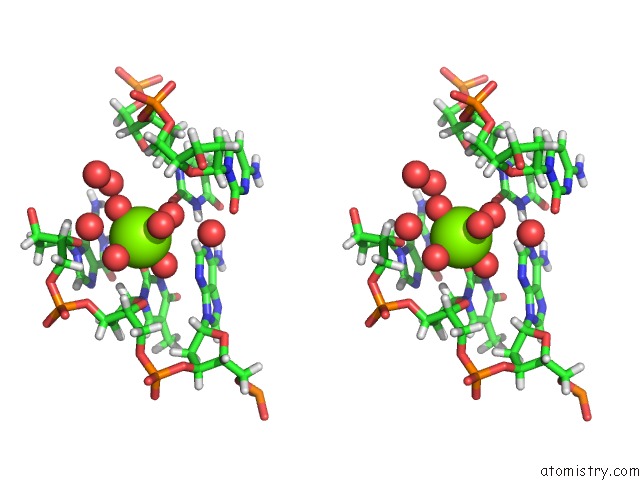
Magnesium binding site 2 out of 4 in 6p0b
Go back to
Magnesium binding site 2 out
of 4 in the Human Dna Ligase 1 (E346A/E592A) Bound to An Adenylated, Dideoxy Terminated Dna Nick with 200 Mm MG2+
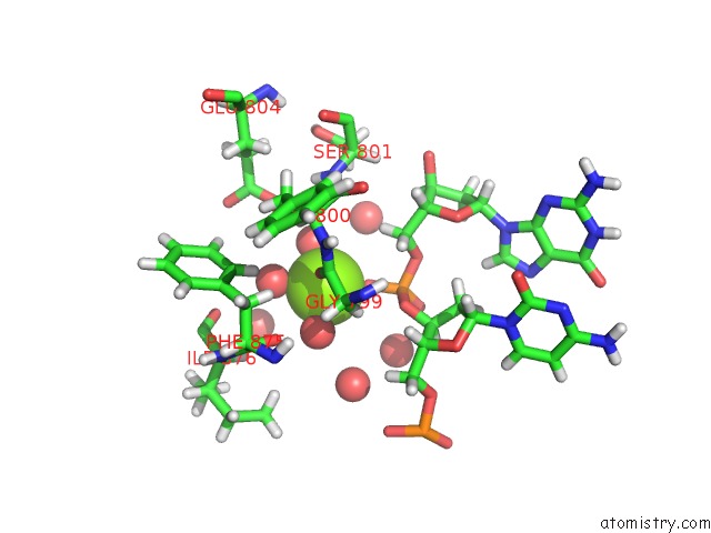
Mono view
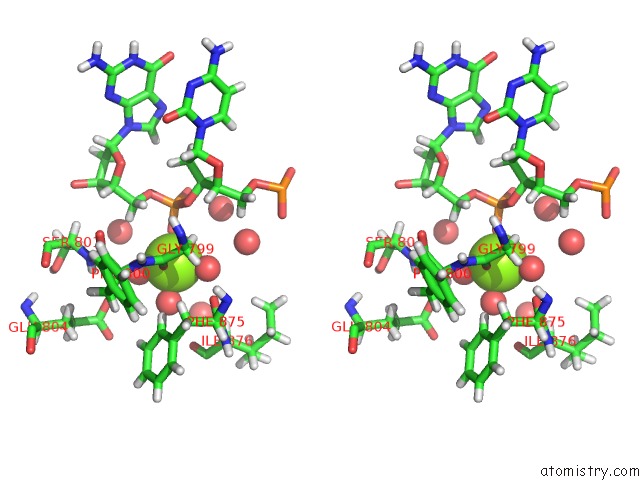
Stereo pair view
Mono view
Stereo pair view
A full contact list of Magnesium with other atoms in the Mg binding
site number 2 of Human Dna Ligase 1 (E346A/E592A) Bound to An Adenylated, Dideoxy Terminated Dna Nick with 200 Mm MG2+ within 5.0Å range:
|
Magnesium binding site 3 out of 4 in 6p0b
Go back to
Magnesium binding site 3 out
of 4 in the Human Dna Ligase 1 (E346A/E592A) Bound to An Adenylated, Dideoxy Terminated Dna Nick with 200 Mm MG2+
Mono view
Stereo pair view
Mono view
Stereo pair view
A full contact list of Magnesium with other atoms in the Mg binding
site number 3 of Human Dna Ligase 1 (E346A/E592A) Bound to An Adenylated, Dideoxy Terminated Dna Nick with 200 Mm MG2+ within 5.0Å range:
|
Magnesium binding site 4 out of 4 in 6p0b
Go back to
Magnesium binding site 4 out
of 4 in the Human Dna Ligase 1 (E346A/E592A) Bound to An Adenylated, Dideoxy Terminated Dna Nick with 200 Mm MG2+
Mono view
Stereo pair view
Mono view
Stereo pair view
A full contact list of Magnesium with other atoms in the Mg binding
site number 4 of Human Dna Ligase 1 (E346A/E592A) Bound to An Adenylated, Dideoxy Terminated Dna Nick with 200 Mm MG2+ within 5.0Å range:
|
Reference:
P.P.Tumbale,
T.J.Jurkiw,
M.J.Schellenberg,
A.A.Riccio,
P.J.O'brien,
R.S.Williams.
Two-Tiered Enforcement of High-Fidelity Dna Ligation. Nat Commun V. 10 5431 2019.
ISSN: ESSN 2041-1723
PubMed: 31780661
DOI: 10.1038/S41467-019-13478-7
Page generated: Tue Oct 1 13:46:05 2024
ISSN: ESSN 2041-1723
PubMed: 31780661
DOI: 10.1038/S41467-019-13478-7
Last articles
Zn in 9J0NZn in 9J0O
Zn in 9J0P
Zn in 9FJX
Zn in 9EKB
Zn in 9C0F
Zn in 9CAH
Zn in 9CH0
Zn in 9CH3
Zn in 9CH1