Magnesium »
PDB 6p1p-6p8a »
6p2g »
Magnesium in PDB 6p2g: Structure of Hiv-1 Reverse Transcriptase (Rt) in Complex with Dsdna and D-Ddctp
Enzymatic activity of Structure of Hiv-1 Reverse Transcriptase (Rt) in Complex with Dsdna and D-Ddctp
All present enzymatic activity of Structure of Hiv-1 Reverse Transcriptase (Rt) in Complex with Dsdna and D-Ddctp:
2.7.7.49; 2.7.7.7; 3.1.26.13;
2.7.7.49; 2.7.7.7; 3.1.26.13;
Protein crystallography data
The structure of Structure of Hiv-1 Reverse Transcriptase (Rt) in Complex with Dsdna and D-Ddctp, PDB code: 6p2g
was solved by
N.Bertoletti,
K.S.Anderson,
with X-Ray Crystallography technique. A brief refinement statistics is given in the table below:
| Resolution Low / High (Å) | 29.59 / 2.99 |
| Space group | C 2 2 21 |
| Cell size a, b, c (Å), α, β, γ (°) | 168.548, 171.578, 106.001, 90.00, 90.00, 90.00 |
| R / Rfree (%) | 21.5 / 26.7 |
Magnesium Binding Sites:
The binding sites of Magnesium atom in the Structure of Hiv-1 Reverse Transcriptase (Rt) in Complex with Dsdna and D-Ddctp
(pdb code 6p2g). This binding sites where shown within
5.0 Angstroms radius around Magnesium atom.
In total only one binding site of Magnesium was determined in the Structure of Hiv-1 Reverse Transcriptase (Rt) in Complex with Dsdna and D-Ddctp, PDB code: 6p2g:
In total only one binding site of Magnesium was determined in the Structure of Hiv-1 Reverse Transcriptase (Rt) in Complex with Dsdna and D-Ddctp, PDB code: 6p2g:
Magnesium binding site 1 out of 1 in 6p2g
Go back to
Magnesium binding site 1 out
of 1 in the Structure of Hiv-1 Reverse Transcriptase (Rt) in Complex with Dsdna and D-Ddctp
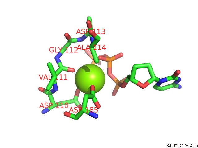
Mono view
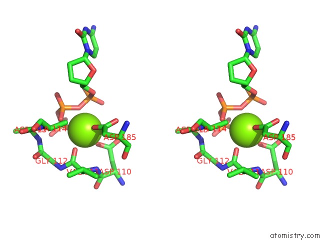
Stereo pair view
Mono view
Stereo pair view
A full contact list of Magnesium with other atoms in the Mg binding
site number 1 of Structure of Hiv-1 Reverse Transcriptase (Rt) in Complex with Dsdna and D-Ddctp within 5.0Å range:
|
Reference:
N.Bertoletti,
A.H.Chan,
R.F.Schinazi,
Y.W.Yin,
K.S.Anderson.
Structural Insights Into the Recognition of Nucleoside Reverse Transcriptase Inhibitors By Hiv-1 Reverse Transcriptase: First Crystal Structures with Reverse Transcriptase and the Active Triphosphate Forms of Lamivudine and Emtricitabine. Protein Sci. V. 28 1664 2019.
ISSN: ESSN 1469-896X
PubMed: 31301259
DOI: 10.1002/PRO.3681
Page generated: Tue Oct 1 13:50:46 2024
ISSN: ESSN 1469-896X
PubMed: 31301259
DOI: 10.1002/PRO.3681
Last articles
Zn in 9J0NZn in 9J0O
Zn in 9J0P
Zn in 9FJX
Zn in 9EKB
Zn in 9C0F
Zn in 9CAH
Zn in 9CH0
Zn in 9CH3
Zn in 9CH1