Magnesium »
PDB 6p1q-6p8b »
6p2h »
Magnesium in PDB 6p2h: Structural Basis For 2'-Deoxyguanosine Recognition By the 2'-Dg-II Class of Riboswitches
Protein crystallography data
The structure of Structural Basis For 2'-Deoxyguanosine Recognition By the 2'-Dg-II Class of Riboswitches, PDB code: 6p2h
was solved by
M.M.Matyjasik,
R.T.Batey,
with X-Ray Crystallography technique. A brief refinement statistics is given in the table below:
| Resolution Low / High (Å) | 29.95 / 2.80 |
| Space group | P 41 2 2 |
| Cell size a, b, c (Å), α, β, γ (°) | 91.805, 91.805, 77.756, 90.00, 90.00, 90.00 |
| R / Rfree (%) | 24.4 / 27.6 |
Other elements in 6p2h:
The structure of Structural Basis For 2'-Deoxyguanosine Recognition By the 2'-Dg-II Class of Riboswitches also contains other interesting chemical elements:
| Cobalt | (Co) | 5 atoms |
Magnesium Binding Sites:
The binding sites of Magnesium atom in the Structural Basis For 2'-Deoxyguanosine Recognition By the 2'-Dg-II Class of Riboswitches
(pdb code 6p2h). This binding sites where shown within
5.0 Angstroms radius around Magnesium atom.
In total 5 binding sites of Magnesium where determined in the Structural Basis For 2'-Deoxyguanosine Recognition By the 2'-Dg-II Class of Riboswitches, PDB code: 6p2h:
Jump to Magnesium binding site number: 1; 2; 3; 4; 5;
In total 5 binding sites of Magnesium where determined in the Structural Basis For 2'-Deoxyguanosine Recognition By the 2'-Dg-II Class of Riboswitches, PDB code: 6p2h:
Jump to Magnesium binding site number: 1; 2; 3; 4; 5;
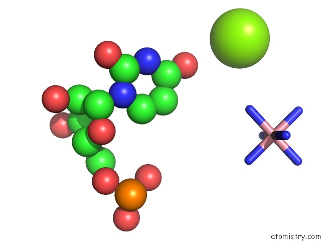
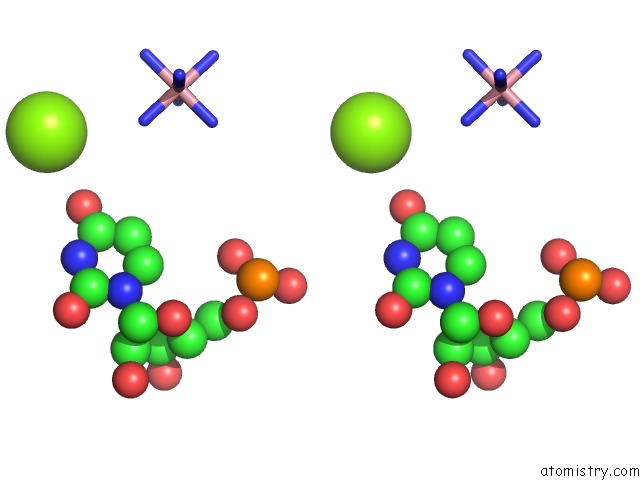
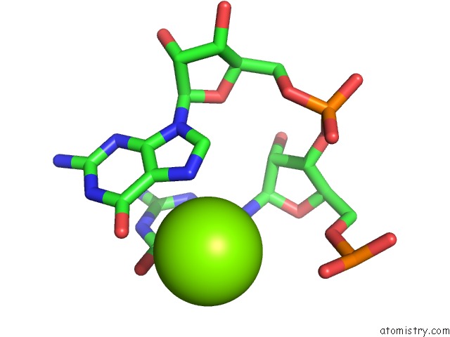
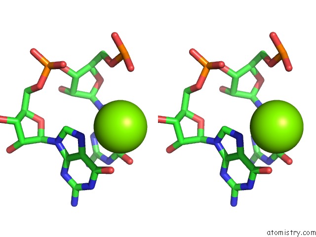
Magnesium binding site 1 out of 5 in 6p2h
Go back to
Magnesium binding site 1 out
of 5 in the Structural Basis For 2'-Deoxyguanosine Recognition By the 2'-Dg-II Class of Riboswitches
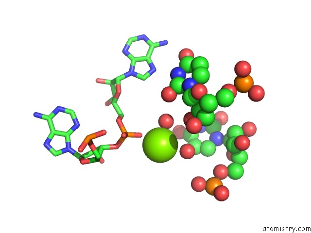
Mono view
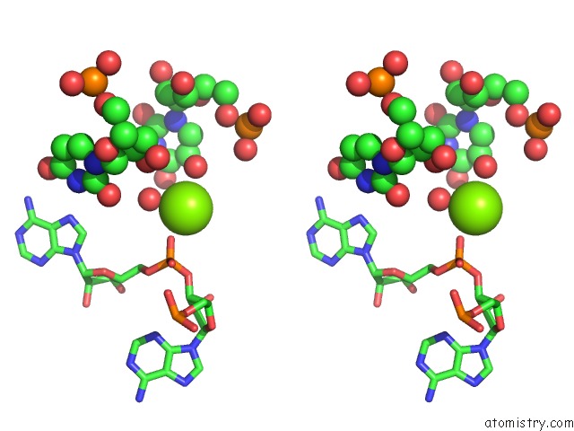
Stereo pair view
Mono view
Stereo pair view
A full contact list of Magnesium with other atoms in the Mg binding
site number 1 of Structural Basis For 2'-Deoxyguanosine Recognition By the 2'-Dg-II Class of Riboswitches within 5.0Å range:
|
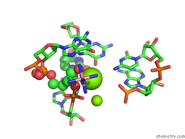
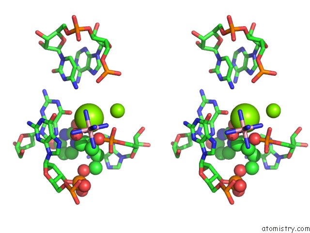
Magnesium binding site 2 out of 5 in 6p2h
Go back to
Magnesium binding site 2 out
of 5 in the Structural Basis For 2'-Deoxyguanosine Recognition By the 2'-Dg-II Class of Riboswitches
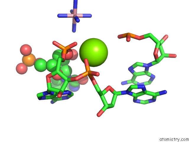
Mono view
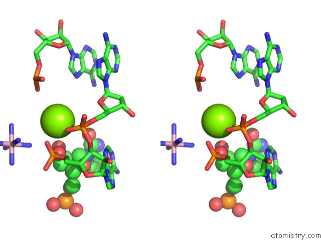
Stereo pair view
Mono view
Stereo pair view
A full contact list of Magnesium with other atoms in the Mg binding
site number 2 of Structural Basis For 2'-Deoxyguanosine Recognition By the 2'-Dg-II Class of Riboswitches within 5.0Å range:
|
Magnesium binding site 3 out of 5 in 6p2h
Go back to
Magnesium binding site 3 out
of 5 in the Structural Basis For 2'-Deoxyguanosine Recognition By the 2'-Dg-II Class of Riboswitches
Mono view
Stereo pair view
Mono view
Stereo pair view
A full contact list of Magnesium with other atoms in the Mg binding
site number 3 of Structural Basis For 2'-Deoxyguanosine Recognition By the 2'-Dg-II Class of Riboswitches within 5.0Å range:
|
Magnesium binding site 4 out of 5 in 6p2h
Go back to
Magnesium binding site 4 out
of 5 in the Structural Basis For 2'-Deoxyguanosine Recognition By the 2'-Dg-II Class of Riboswitches
Mono view
Stereo pair view
Mono view
Stereo pair view
A full contact list of Magnesium with other atoms in the Mg binding
site number 4 of Structural Basis For 2'-Deoxyguanosine Recognition By the 2'-Dg-II Class of Riboswitches within 5.0Å range:
|
Magnesium binding site 5 out of 5 in 6p2h
Go back to
Magnesium binding site 5 out
of 5 in the Structural Basis For 2'-Deoxyguanosine Recognition By the 2'-Dg-II Class of Riboswitches
Mono view
Stereo pair view
Mono view
Stereo pair view
A full contact list of Magnesium with other atoms in the Mg binding
site number 5 of Structural Basis For 2'-Deoxyguanosine Recognition By the 2'-Dg-II Class of Riboswitches within 5.0Å range:
|
Reference:
M.M.Matyjasik,
R.T.Batey.
Structural Basis For 2'-Deoxyguanosine Recognition By the 2'-Dg-II Class of Riboswitches. Nucleic Acids Res. V. 47 10931 2019.
ISSN: ESSN 1362-4962
PubMed: 31598729
DOI: 10.1093/NAR/GKZ839
Page generated: Tue Oct 1 13:50:48 2024
ISSN: ESSN 1362-4962
PubMed: 31598729
DOI: 10.1093/NAR/GKZ839
Last articles
F in 4IVMF in 4IV2
F in 4IUI
F in 4IN4
F in 4IU7
F in 4ITI
F in 4IUE
F in 4IRU
F in 4ITJ
F in 4ISF