Magnesium »
PDB 6p1o-6p89 »
6p85 »
Magnesium in PDB 6p85: E.Coli Lpxd in Complex with Compound 3
Enzymatic activity of E.Coli Lpxd in Complex with Compound 3
All present enzymatic activity of E.Coli Lpxd in Complex with Compound 3:
2.3.1.191;
2.3.1.191;
Protein crystallography data
The structure of E.Coli Lpxd in Complex with Compound 3, PDB code: 6p85
was solved by
X.Ma,
S.Shia,
with X-Ray Crystallography technique. A brief refinement statistics is given in the table below:
| Resolution Low / High (Å) | 42.83 / 1.90 |
| Space group | P 32 2 1 |
| Cell size a, b, c (Å), α, β, γ (°) | 98.911, 98.911, 215.312, 90.00, 90.00, 120.00 |
| R / Rfree (%) | 17 / 19.9 |
Magnesium Binding Sites:
The binding sites of Magnesium atom in the E.Coli Lpxd in Complex with Compound 3
(pdb code 6p85). This binding sites where shown within
5.0 Angstroms radius around Magnesium atom.
In total 2 binding sites of Magnesium where determined in the E.Coli Lpxd in Complex with Compound 3, PDB code: 6p85:
Jump to Magnesium binding site number: 1; 2;
In total 2 binding sites of Magnesium where determined in the E.Coli Lpxd in Complex with Compound 3, PDB code: 6p85:
Jump to Magnesium binding site number: 1; 2;
Magnesium binding site 1 out of 2 in 6p85
Go back to
Magnesium binding site 1 out
of 2 in the E.Coli Lpxd in Complex with Compound 3
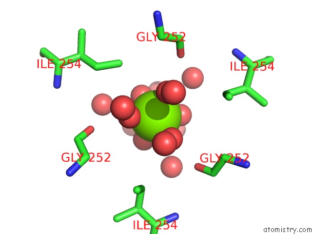
Mono view
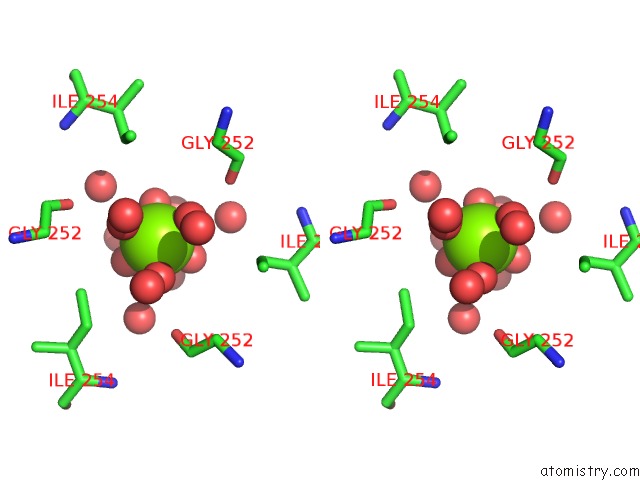
Stereo pair view
Mono view
Stereo pair view
A full contact list of Magnesium with other atoms in the Mg binding
site number 1 of E.Coli Lpxd in Complex with Compound 3 within 5.0Å range:
|
Magnesium binding site 2 out of 2 in 6p85
Go back to
Magnesium binding site 2 out
of 2 in the E.Coli Lpxd in Complex with Compound 3
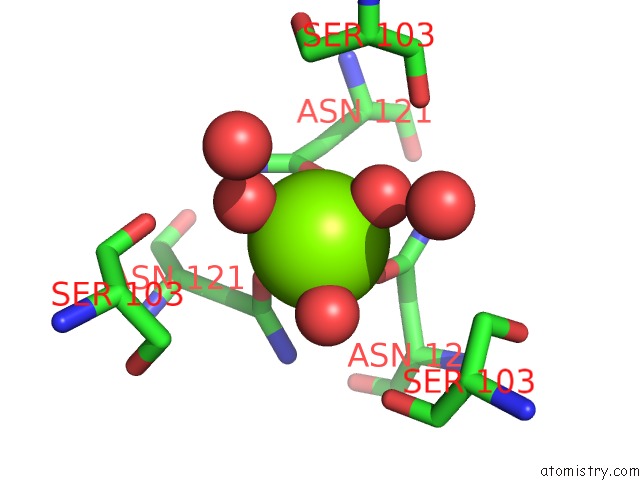
Mono view
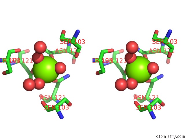
Stereo pair view
Mono view
Stereo pair view
A full contact list of Magnesium with other atoms in the Mg binding
site number 2 of E.Coli Lpxd in Complex with Compound 3 within 5.0Å range:
|
Reference:
X.Ma,
R.Prathapam,
C.Wartchow,
B.Chie Leon,
C.M.Ho,
J.De Vicente,
W.Han,
M.Li,
Y.Lu,
S.Ramurthy,
S.Shia,
M.Steffek,
T.Uehara.
Structural and Biological Basis of Small Molecule Inhibition of Escherichia Coli Lpxd Acyltransferase Essential For Lipopolysaccharide Biosynthesis. Acs Infect Dis. 2019.
ISSN: ESSN 2373-8227
PubMed: 31402665
DOI: 10.1021/ACSINFECDIS.9B00127
Page generated: Tue Oct 1 13:55:52 2024
ISSN: ESSN 2373-8227
PubMed: 31402665
DOI: 10.1021/ACSINFECDIS.9B00127
Last articles
Zn in 9JYWZn in 9IR4
Zn in 9IR3
Zn in 9GMX
Zn in 9GMW
Zn in 9JEJ
Zn in 9ERF
Zn in 9ERE
Zn in 9EGV
Zn in 9EGW