Magnesium »
PDB 6p8b-6pek »
6pcl »
Magnesium in PDB 6pcl: Crystal Structure of Human Diphosphoinositol Polyphosphate Phosphohydrolase 1 in Complex with 5-IP7
Enzymatic activity of Crystal Structure of Human Diphosphoinositol Polyphosphate Phosphohydrolase 1 in Complex with 5-IP7
All present enzymatic activity of Crystal Structure of Human Diphosphoinositol Polyphosphate Phosphohydrolase 1 in Complex with 5-IP7:
3.6.1.52;
3.6.1.52;
Protein crystallography data
The structure of Crystal Structure of Human Diphosphoinositol Polyphosphate Phosphohydrolase 1 in Complex with 5-IP7, PDB code: 6pcl
was solved by
D.E.Dollins,
J.Neubauer,
J.Dong,
J.D.York,
with X-Ray Crystallography technique. A brief refinement statistics is given in the table below:
| Resolution Low / High (Å) | 27.60 / 1.30 |
| Space group | P 21 21 21 |
| Cell size a, b, c (Å), α, β, γ (°) | 45.560, 59.576, 62.358, 90.00, 90.00, 90.00 |
| R / Rfree (%) | 14.3 / 16.1 |
Other elements in 6pcl:
The structure of Crystal Structure of Human Diphosphoinositol Polyphosphate Phosphohydrolase 1 in Complex with 5-IP7 also contains other interesting chemical elements:
| Chlorine | (Cl) | 2 atoms |
Magnesium Binding Sites:
The binding sites of Magnesium atom in the Crystal Structure of Human Diphosphoinositol Polyphosphate Phosphohydrolase 1 in Complex with 5-IP7
(pdb code 6pcl). This binding sites where shown within
5.0 Angstroms radius around Magnesium atom.
In total 3 binding sites of Magnesium where determined in the Crystal Structure of Human Diphosphoinositol Polyphosphate Phosphohydrolase 1 in Complex with 5-IP7, PDB code: 6pcl:
Jump to Magnesium binding site number: 1; 2; 3;
In total 3 binding sites of Magnesium where determined in the Crystal Structure of Human Diphosphoinositol Polyphosphate Phosphohydrolase 1 in Complex with 5-IP7, PDB code: 6pcl:
Jump to Magnesium binding site number: 1; 2; 3;
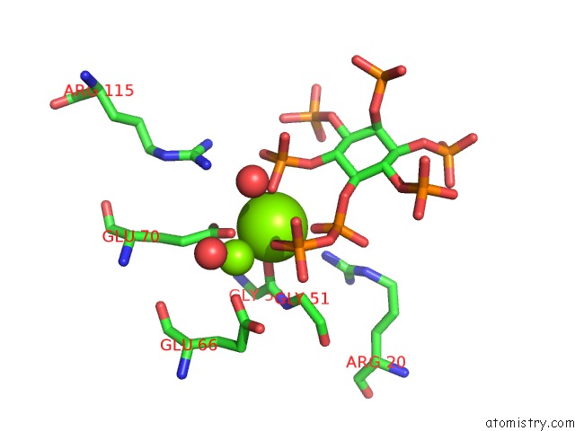
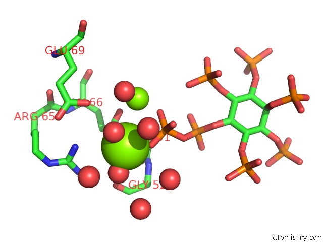
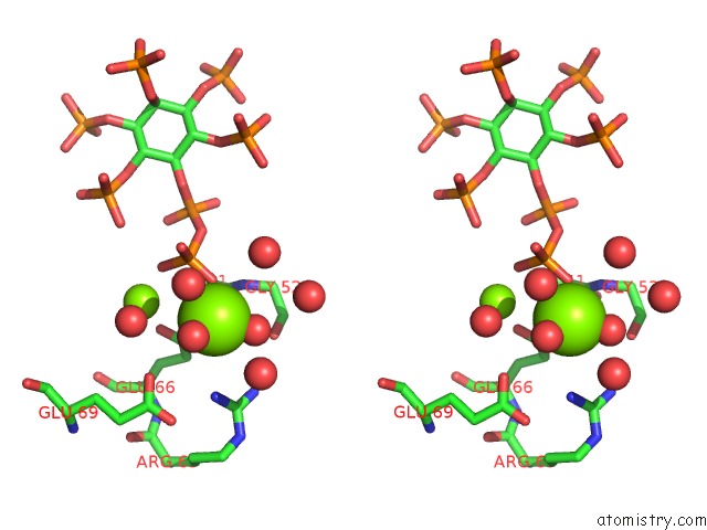
Magnesium binding site 1 out of 3 in 6pcl
Go back to
Magnesium binding site 1 out
of 3 in the Crystal Structure of Human Diphosphoinositol Polyphosphate Phosphohydrolase 1 in Complex with 5-IP7
Mono view
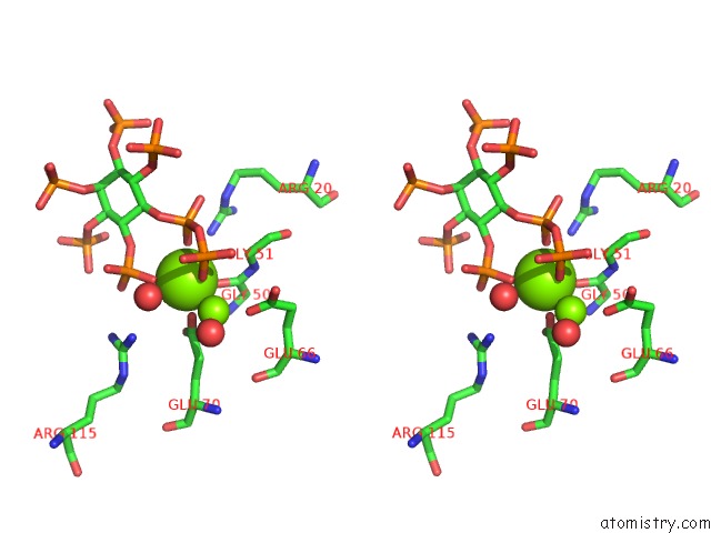
Stereo pair view
Mono view
Stereo pair view
A full contact list of Magnesium with other atoms in the Mg binding
site number 1 of Crystal Structure of Human Diphosphoinositol Polyphosphate Phosphohydrolase 1 in Complex with 5-IP7 within 5.0Å range:
|
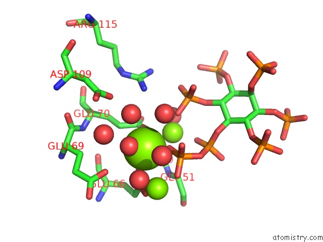
Magnesium binding site 2 out of 3 in 6pcl
Go back to
Magnesium binding site 2 out
of 3 in the Crystal Structure of Human Diphosphoinositol Polyphosphate Phosphohydrolase 1 in Complex with 5-IP7
Mono view
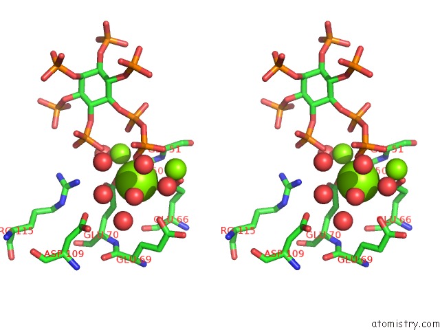
Stereo pair view
Mono view
Stereo pair view
A full contact list of Magnesium with other atoms in the Mg binding
site number 2 of Crystal Structure of Human Diphosphoinositol Polyphosphate Phosphohydrolase 1 in Complex with 5-IP7 within 5.0Å range:
|
Magnesium binding site 3 out of 3 in 6pcl
Go back to
Magnesium binding site 3 out
of 3 in the Crystal Structure of Human Diphosphoinositol Polyphosphate Phosphohydrolase 1 in Complex with 5-IP7
Mono view
Stereo pair view
Mono view
Stereo pair view
A full contact list of Magnesium with other atoms in the Mg binding
site number 3 of Crystal Structure of Human Diphosphoinositol Polyphosphate Phosphohydrolase 1 in Complex with 5-IP7 within 5.0Å range:
|
Reference:
D.E.Dollins,
W.Bai,
P.C.Fridy,
J.C.Otto,
J.L.Neubauer,
S.G.Gattis,
K.P.M.Mehta,
J.D.York.
VIP1 Is A Kinase and Pyrophosphatase Switch That Regulates Inositol Diphosphate Signaling. Proc.Natl.Acad.Sci.Usa 2020.
ISSN: ESSN 1091-6490
PubMed: 32303658
DOI: 10.1073/PNAS.1908875117
Page generated: Tue Oct 1 14:03:27 2024
ISSN: ESSN 1091-6490
PubMed: 32303658
DOI: 10.1073/PNAS.1908875117
Last articles
Zn in 9J0NZn in 9J0O
Zn in 9J0P
Zn in 9FJX
Zn in 9EKB
Zn in 9C0F
Zn in 9CAH
Zn in 9CH0
Zn in 9CH3
Zn in 9CH1