Magnesium in PDB 6peu: Structure of Ycao Enzyme From Methanocaldococcus Jannaschii in Complex with Peptide

Protein crystallography data

The structure of Structure of Ycao Enzyme From Methanocaldococcus Jannaschii in Complex with Peptide, PDB code: 6peu was solved by S.-H.Dong, S.K.Nair, with X-Ray Crystallography technique. A brief refinement statistics is given in the table below:

Resolution Low / High (Å) 25.00 / 1.95
Space group P 1 21 1
Cell size a, b, c (Å), α, β, γ (°) 59.324, 149.755, 105.671, 90.00, 103.57, 90.00
R / Rfree (%) 19.5 / 23.7

Magnesium Binding Sites:

The binding sites of Magnesium atom in the Structure of Ycao Enzyme From Methanocaldococcus Jannaschii in Complex with Peptide (pdb code 6peu). This binding sites where shown within 5.0 Angstroms radius around Magnesium atom.
In total 7 binding sites of Magnesium where determined in the Structure of Ycao Enzyme From Methanocaldococcus Jannaschii in Complex with Peptide, PDB code: 6peu:
Jump to Magnesium binding site number: 1; 2; 3; 4; 5; 6; 7;

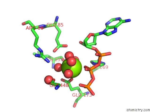
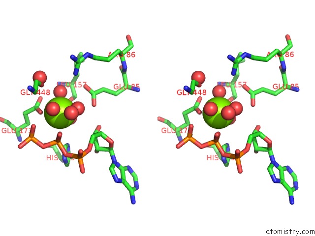
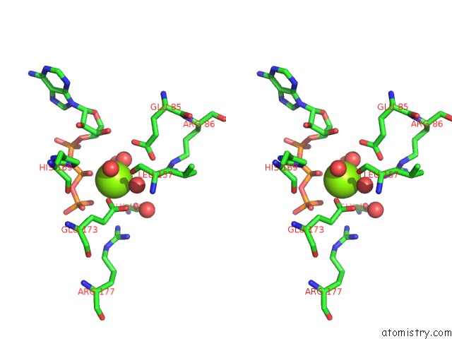
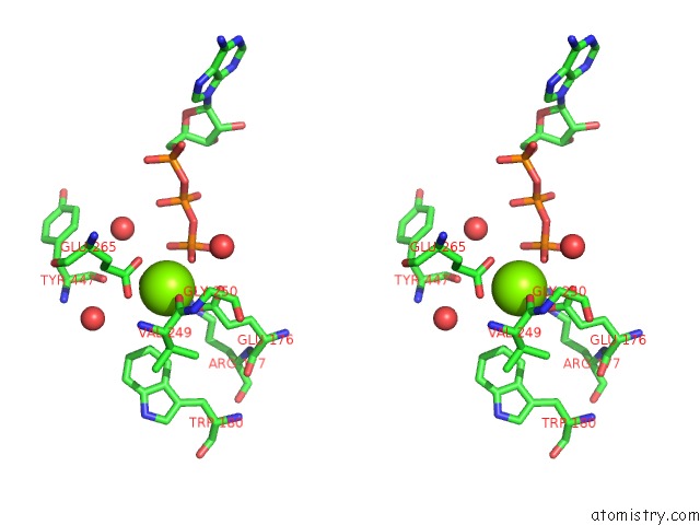
Magnesium binding site 1 out of 7 in 6peu

Go back to Magnesium Binding Sites List in 6peu
Magnesium binding site 1 out of 7 in the Structure of Ycao Enzyme From Methanocaldococcus Jannaschii in Complex with Peptide


Mono view


Stereo pair view

A full contact list of Magnesium with other atoms in the Mg binding site number 1 of Structure of Ycao Enzyme From Methanocaldococcus Jannaschii in Complex with Peptide within 5.0Å range:
probe atom residue distance (Å) B Occ
A:Mg501

b:33.8
occ:1.00
O A:HOH690 2.2 29.2 1.0
OE2 A:GLU173 2.2 27.0 1.0
O M:HOH501 2.2 25.9 1.0
O1B A:ATP500 2.3 34.8 1.0
O A:HOH620 2.3 26.7 1.0
O A:HOH645 2.4 23.6 1.0
CD A:GLU173 3.3 25.7 1.0
PB A:ATP500 3.5 37.0 1.0
OE1 A:GLU173 3.6 24.4 1.0
O A:HOH601 3.8 32.4 1.0
O2B A:ATP500 3.8 36.1 1.0
O1G A:ATP500 3.8 34.1 1.0
O2G A:ATP500 3.9 34.1 1.0
OE1 A:GLU85 3.9 27.6 1.0
O A:LEU157 4.1 23.5 1.0
NE2 A:HIS169 4.2 31.8 1.0
OE2 A:GLU85 4.2 27.4 1.0
O3B A:ATP500 4.2 36.1 1.0
NH2 A:ARG86 4.2 23.1 1.0
PG A:ATP500 4.3 35.5 1.0
O M:HOH502 4.3 24.8 1.0
O M:GLY448 4.3 30.8 1.0
CD A:GLU85 4.5 27.5 1.0
C3' A:ATP500 4.6 24.4 1.0
CG A:GLU173 4.6 24.2 1.0
C5' A:ATP500 4.6 27.1 1.0
CD2 A:HIS169 4.6 30.5 1.0
O3A A:ATP500 4.8 35.2 1.0
C4' A:ATP500 4.9 26.2 1.0
O3' A:ATP500 4.9 23.3 1.0

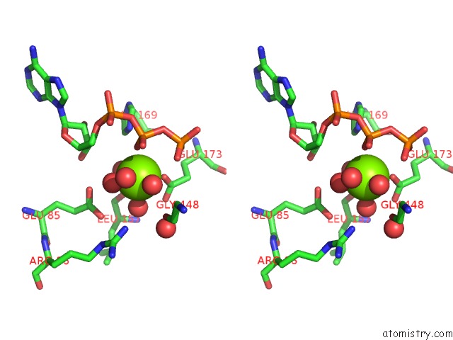
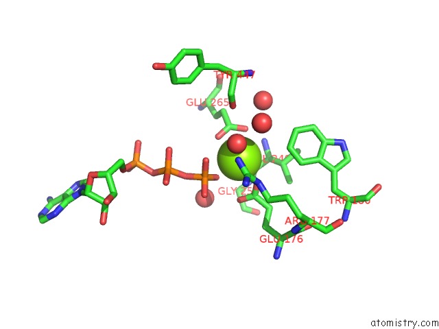
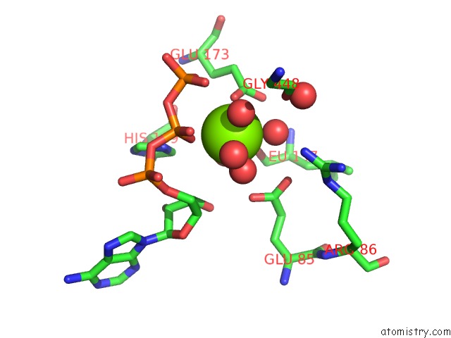
Magnesium binding site 2 out of 7 in 6peu

Go back to Magnesium Binding Sites List in 6peu
Magnesium binding site 2 out of 7 in the Structure of Ycao Enzyme From Methanocaldococcus Jannaschii in Complex with Peptide


Mono view


Stereo pair view

A full contact list of Magnesium with other atoms in the Mg binding site number 2 of Structure of Ycao Enzyme From Methanocaldococcus Jannaschii in Complex with Peptide within 5.0Å range:
probe atom residue distance (Å) B Occ
A:Mg502

b:29.1
occ:1.00
O A:HOH657 2.1 34.8 1.0
O3G A:ATP500 2.3 32.3 1.0
O A:VAL249 2.4 25.6 1.0
OE2 A:GLU176 2.4 25.2 1.0
OE2 A:GLU265 2.5 25.5 1.0
OE1 A:GLU265 2.7 24.1 1.0
CD A:GLU265 2.9 24.6 1.0
CD A:GLU176 3.1 26.3 1.0
PG A:ATP500 3.2 35.5 1.0
O1G A:ATP500 3.3 34.1 1.0
OE1 A:GLU176 3.3 26.8 1.0
C A:VAL249 3.5 24.6 1.0
O A:HOH746 4.0 48.8 1.0
CA A:VAL249 4.1 25.1 1.0
N A:VAL249 4.1 25.0 1.0
CB A:VAL249 4.1 25.4 1.0
O2G A:ATP500 4.2 34.1 1.0
CZ3 A:TRP180 4.3 28.5 1.0
CG A:GLU265 4.4 23.6 1.0
O M:HOH506 4.4 0.2 1.0
O3B A:ATP500 4.4 36.1 1.0
NH2 A:ARG177 4.4 24.2 1.0
O M:TYR447 4.4 29.5 1.0
NE A:ARG177 4.5 24.7 1.0
CG A:GLU176 4.5 24.8 1.0
O A:HOH738 4.6 35.7 1.0
N A:GLY250 4.7 23.5 1.0
O A:HOH653 4.9 30.3 1.0
CZ A:ARG177 4.9 24.5 1.0
CG1 A:VAL249 4.9 26.0 1.0

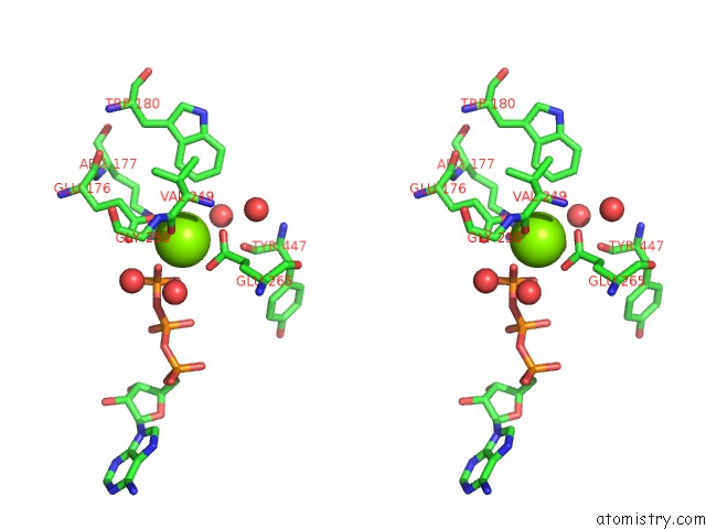
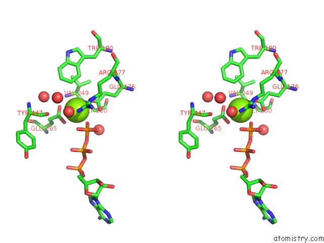
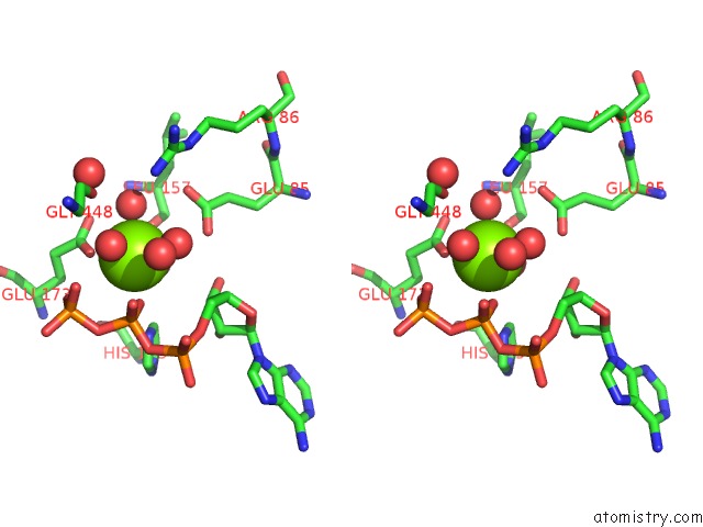
Magnesium binding site 3 out of 7 in 6peu

Go back to Magnesium Binding Sites List in 6peu
Magnesium binding site 3 out of 7 in the Structure of Ycao Enzyme From Methanocaldococcus Jannaschii in Complex with Peptide


Mono view


Stereo pair view

A full contact list of Magnesium with other atoms in the Mg binding site number 3 of Structure of Ycao Enzyme From Methanocaldococcus Jannaschii in Complex with Peptide within 5.0Å range:
probe atom residue distance (Å) B Occ
B:Mg501

b:29.9
occ:1.00
O B:HOH740 2.1 25.4 1.0
O B:HOH688 2.2 29.3 1.0
O B:HOH634 2.3 23.8 1.0
O1B B:ATP500 2.3 33.2 1.0
OE2 B:GLU173 2.3 21.4 1.0
O B:HOH670 2.5 24.2 1.0
CD B:GLU173 3.3 20.4 1.0
PB B:ATP500 3.5 39.6 1.0
OE1 B:GLU173 3.7 21.2 1.0
O B:HOH615 3.8 32.9 1.0
O2G B:ATP500 3.8 39.4 1.0
O2B B:ATP500 3.8 36.9 1.0
O1G B:ATP500 3.9 38.8 1.0
OE1 B:GLU85 4.0 21.4 1.0
O B:LEU157 4.1 21.0 1.0
CE1 B:HIS169 4.2 25.1 1.0
OE2 B:GLU85 4.2 22.0 1.0
PG B:ATP500 4.3 42.4 1.0
O3B B:ATP500 4.3 39.8 1.0
O N:GLY448 4.3 23.1 1.0
O N:HOH501 4.4 19.9 1.0
CD B:GLU85 4.5 21.0 1.0
C3' B:ATP500 4.6 23.1 1.0
C5' B:ATP500 4.6 24.6 1.0
CG B:GLU173 4.6 19.6 1.0
NH2 B:ARG86 4.7 33.5 1.0
ND1 B:HIS169 4.8 25.4 1.0
O3A B:ATP500 4.8 33.7 1.0
C4' B:ATP500 4.9 23.8 1.0
O3' B:ATP500 4.9 22.0 1.0

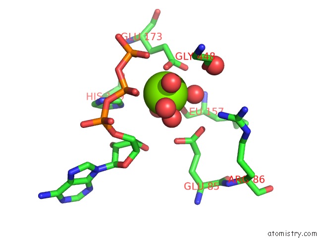
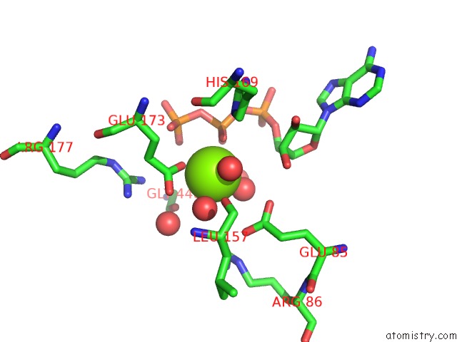
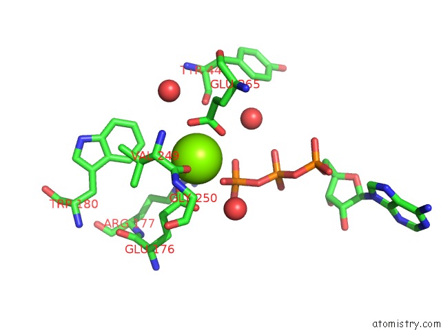
Magnesium binding site 4 out of 7 in 6peu

Go back to Magnesium Binding Sites List in 6peu
Magnesium binding site 4 out of 7 in the Structure of Ycao Enzyme From Methanocaldococcus Jannaschii in Complex with Peptide


Mono view


Stereo pair view

A full contact list of Magnesium with other atoms in the Mg binding site number 4 of Structure of Ycao Enzyme From Methanocaldococcus Jannaschii in Complex with Peptide within 5.0Å range:
probe atom residue distance (Å) B Occ
B:Mg502

b:34.3
occ:1.00
O B:HOH642 2.3 32.9 1.0
O3G B:ATP500 2.3 39.2 1.0
O B:VAL249 2.4 25.6 1.0
OE2 B:GLU176 2.4 22.8 1.0
OE2 B:GLU265 2.5 23.9 1.0
OE1 B:GLU265 2.6 23.3 1.0
CD B:GLU265 2.9 24.2 1.0
CD B:GLU176 3.1 23.3 1.0
OE1 B:GLU176 3.2 25.5 1.0
O1G B:ATP500 3.2 38.8 1.0
PG B:ATP500 3.2 42.4 1.0
C B:VAL249 3.5 23.9 1.0
N B:VAL249 4.0 25.5 1.0
CA B:VAL249 4.0 24.2 1.0
CB B:VAL249 4.1 24.5 1.0
CZ3 B:TRP180 4.2 23.8 1.0
O2G B:ATP500 4.3 39.4 1.0
CG B:GLU265 4.4 22.1 1.0
O3B B:ATP500 4.4 39.8 1.0
O N:TYR447 4.5 26.3 1.0
CG B:GLU176 4.5 23.1 1.0
O N:HOH503 4.5 0.2 1.0
NE B:ARG177 4.5 21.0 1.0
NH2 B:ARG177 4.5 21.7 1.0
N B:GLY250 4.6 22.3 1.0
O B:HOH757 4.7 53.2 1.0
O B:HOH672 4.9 27.2 1.0
CE3 B:TRP180 4.9 23.6 1.0
CH2 B:TRP180 5.0 23.8 1.0
CG1 B:VAL249 5.0 23.9 1.0

Magnesium binding site 5 out of 7 in 6peu

Go back to Magnesium Binding Sites List in 6peu
Magnesium binding site 5 out of 7 in the Structure of Ycao Enzyme From Methanocaldococcus Jannaschii in Complex with Peptide


Mono view


Stereo pair view

A full contact list of Magnesium with other atoms in the Mg binding site number 5 of Structure of Ycao Enzyme From Methanocaldococcus Jannaschii in Complex with Peptide within 5.0Å range:
probe atom residue distance (Å) B Occ
C:Mg501

b:31.2
occ:1.00
O C:HOH642 2.1 29.5 1.0
OE2 C:GLU173 2.2 25.6 1.0
O C:HOH672 2.3 24.4 1.0
O1B C:ATP500 2.4 30.9 1.0
O C:HOH624 2.4 24.0 1.0
O P:HOH501 2.5 29.0 1.0
CD C:GLU173 3.2 24.9 1.0
PB C:ATP500 3.5 39.6 1.0
OE1 C:GLU173 3.6 24.2 1.0
O2B C:ATP500 3.8 38.4 1.0
O C:HOH616 3.8 42.4 1.0
O2G C:ATP500 3.9 36.9 1.0
OE1 C:GLU85 4.0 27.5 1.0
O1G C:ATP500 4.0 38.7 1.0
O C:LEU157 4.0 21.7 1.0
O P:HOH503 4.1 21.5 1.0
OE2 C:GLU85 4.2 27.6 1.0
NE2 C:HIS169 4.2 28.0 1.0
NH2 C:ARG86 4.3 22.7 1.0
O P:GLY448 4.3 26.5 1.0
O3B C:ATP500 4.3 39.7 1.0
PG C:ATP500 4.3 40.6 1.0
CD C:GLU85 4.5 27.7 1.0
CG C:GLU173 4.5 24.8 1.0
C3' C:ATP500 4.6 23.1 1.0
CD2 C:HIS169 4.7 26.6 1.0
C5' C:ATP500 4.7 24.5 1.0
O3' C:ATP500 4.8 24.0 1.0
O3A C:ATP500 4.8 33.7 1.0
NH2 C:ARG177 4.9 26.9 1.0

Magnesium binding site 6 out of 7 in 6peu

Go back to Magnesium Binding Sites List in 6peu
Magnesium binding site 6 out of 7 in the Structure of Ycao Enzyme From Methanocaldococcus Jannaschii in Complex with Peptide


Mono view


Stereo pair view

A full contact list of Magnesium with other atoms in the Mg binding site number 6 of Structure of Ycao Enzyme From Methanocaldococcus Jannaschii in Complex with Peptide within 5.0Å range:
probe atom residue distance (Å) B Occ
D:Mg501

b:36.2
occ:1.00
O D:HOH638 2.1 26.3 1.0
O D:HOH657 2.3 32.4 1.0
OE2 D:GLU173 2.3 25.5 1.0
O1B D:ATP500 2.3 37.4 1.0
O D:HOH609 2.5 35.7 1.0
O D:HOH634 2.6 25.9 1.0
CD D:GLU173 3.3 24.5 1.0
PB D:ATP500 3.4 43.0 1.0
OE1 D:GLU173 3.7 25.3 1.0
O2B D:ATP500 3.7 43.1 1.0
O2G D:ATP500 3.8 36.4 1.0
O D:HOH618 3.9 32.6 1.0
OE1 D:GLU85 3.9 29.1 1.0
O1G D:ATP500 3.9 39.3 1.0
O D:LEU157 4.1 27.4 1.0
O3B D:ATP500 4.2 40.0 1.0
PG D:ATP500 4.2 37.3 1.0
OE2 D:GLU85 4.3 31.4 1.0
NE2 D:HIS169 4.3 33.6 1.0
NH2 D:ARG86 4.4 25.6 1.0
C3' D:ATP500 4.4 27.4 1.0
O Q:GLY448 4.4 29.3 1.0
O Q:HOH501 4.5 21.6 1.0
CD D:GLU85 4.5 30.0 1.0
C5' D:ATP500 4.6 28.3 1.0
CD2 D:HIS169 4.6 31.4 1.0
CG D:GLU173 4.6 24.8 1.0
O3' D:ATP500 4.7 28.1 1.0
C4' D:ATP500 4.8 29.1 1.0
O3A D:ATP500 4.8 40.4 1.0
O5' D:ATP500 5.0 29.8 1.0

Magnesium binding site 7 out of 7 in 6peu

Go back to Magnesium Binding Sites List in 6peu
Magnesium binding site 7 out of 7 in the Structure of Ycao Enzyme From Methanocaldococcus Jannaschii in Complex with Peptide


Mono view


Stereo pair view

A full contact list of Magnesium with other atoms in the Mg binding site number 7 of Structure of Ycao Enzyme From Methanocaldococcus Jannaschii in Complex with Peptide within 5.0Å range:
probe atom residue distance (Å) B Occ
D:Mg502

b:34.4
occ:1.00
O D:HOH631 2.2 32.9 1.0
O D:VAL249 2.3 25.6 1.0
O3G D:ATP500 2.4 35.6 1.0
OE2 D:GLU176 2.4 28.0 1.0
OE1 D:GLU265 2.5 26.1 1.0
OE2 D:GLU265 2.6 25.6 1.0
CD D:GLU265 2.9 24.2 1.0
CD D:GLU176 3.1 27.8 1.0
O1G D:ATP500 3.3 39.3 1.0
OE1 D:GLU176 3.3 29.1 1.0
PG D:ATP500 3.3 37.3 1.0
C D:VAL249 3.4 25.5 1.0
N D:VAL249 4.0 24.4 1.0
CA D:VAL249 4.0 25.8 1.0
CB D:VAL249 4.1 25.4 1.0
O2G D:ATP500 4.3 36.4 1.0
CG D:GLU265 4.4 23.8 1.0
CZ3 D:TRP180 4.4 26.0 1.0
CG D:GLU176 4.5 26.9 1.0
O3B D:ATP500 4.5 40.0 1.0
N D:GLY250 4.5 26.8 1.0
O Q:TYR447 4.5 29.4 1.0
NE D:ARG177 4.6 23.6 1.0
NH2 D:ARG177 4.6 21.9 1.0
O D:HOH735 4.7 35.8 1.0
CG1 D:VAL249 4.8 26.1 1.0
O D:HOH616 4.8 48.4 1.0
CA D:GLY250 4.9 26.4 1.0
O D:HOH711 5.0 32.7 1.0

Reference:

S.H.Dong, A.Liu, N.Mahanta, D.A.Mitchell, S.K.Nair. Mechanistic Basis For Ribosomal Peptide Backbone Modifications. Acs Cent.Sci. V. 5 842 2019.
ISSN: ESSN 2374-7951
PubMed: 31139720
DOI: 10.1021/ACSCENTSCI.9B00124
Page generated: Mon Dec 14 23:47:45 2020

Last articles

Zn in 8WB0
Zn in 8WAX
Zn in 8WAU
Zn in 8WAZ
Zn in 8WAY
Zn in 8WAV
Zn in 8WAW
Zn in 8WAT
Zn in 8W7M
Zn in 8WD3
© Copyright 2008-2020 by atomistry.com
Home   |    Site Map   |    Copyright   |    Contact us   |    Privacy